VENTRICLES OF THE BRAIN (ventriculi cerebri) - cavities located in the brain, lined with ependyma and filled with cerebrospinal fluid. The functional significance of gastrointestinal tracts is determined by the fact that they are the site of formation and reservoir of cerebrospinal fluid (see), as well as part of the cerebrospinal fluid ducts.
Available four ventricles: lateral ventricles(ventriculi lat., first and second), third ventricle(ventriculus tertius) and fourth ventricle(ventriculus quartus). First described by Herophilus in the 4th century. BC e. The discoveries of the cerebral aqueduct by F. Sylvius, the interventricular foramen by A. Monroe, the median opening of the fourth ventricle by F. Magendie, the lateral openings of the fourth ventricle by G. Lushka, as well as the introduction into medicine were of great importance in the study of liquor-conducting tracts. practice of the ventriculography method by W. Dendy (1918).
The forward movement of cerebrospinal fluid is directed from the gland through the unpaired median opening of the fourth ventricle (Magendie) and the paired lateral openings of the fourth ventricle (Lushka) into the cerebellar cistern, from there the cerebrospinal fluid spreads through the cisterns of the base of the brain, channels along the convolutions of the brain onto its convex surface and into the subarachnoid space of the spinal cord and its central canal. The capacity of all ventricles is 30-50 ml.
Embryology
The gastrointestinal tract, as well as the cavities of the spinal cord [the central canal (canalis centralis) and the terminal ventricle (ventriculus terminalis)], are formed as a result of transformations of the primary cavity of the neural tube - the neural canal. The nerve canal along the spinal cord gradually narrows and turns into the central canal and the terminal ventricle. The anterior end of the neural tube expands and then dismembers, forming at the 4th week. development of three brain vesicles (Fig. 1): anterior, middle and rhomboid. At 5-6 weeks. development by differentiation of three brain vesicles, five vesicles are formed, giving rise to five main parts of the brain: telencephalon, diencephalon, midbrain (mesencephalon), hindbrain (metencephalon), medulla oblongata (myelencephalon).
The telencephalon rapidly grows to the sides, forming two lateral bubbles - the rudiments of the hemispheres big brain. The primary cavity of the telencephalon (telocele) gives rise to the cavities of the lateral vesicles, which represent the anlage of the lateral ventricles. At 6-7 weeks. development, the growth of the lateral bladders occurs in the lateral and anterior directions, which leads to the formation of the anterior horn of the lateral ventricles; at 8-10 weeks. growth of the lateral vesicles in the opposite direction is observed, as a result of which the posterior and lower horns of the ventricles appear. Due to the increased growth of the temporal lobes of the brain, the inferior horns of the ventricles move laterally, downward and forward. The part of the cavity of the telencephalon, located in connection with the cavities of the lateral vesicles, turns into interventricular foramina (foramina interventricularia), which communicate the lateral ventricles with the anterior part of the third ventricle. The primary cavity of the diencephalon (diocele) narrows, maintaining a connection with the middle part of the telencephalon cavity, and gives rise to the third ventricle. The cavity of the midbrain (mesocele), which passes in front into the third ventricle, narrows very strongly even at the 7th week. turns into a narrow channel - the cerebral aqueduct (aqueductus cerebri), connecting the third ventricle with the fourth. At the same time, the cavity of the rhombencephalon, which gives rise to the hindbrain and medulla oblongata, expanding laterally, forms the fourth ventricle with its lateral recesses (recessus lat.). The vascular base of the fourth ventricle (tela chorioidea ventriculi quarti) initially almost completely closes its cavity (with the exception of the opening of the cerebral aqueduct). By the 10th week. During development, openings are formed in it and in the wall of the ventricle: one median (apertura mediana) at the lower corner of the rhomboid fossa and two paired lateral ones (aperturae lat.) at the tops of the lateral recesses. Through these openings, the fourth ventricle communicates with the subarachnoid space of the brain. The cavity of the fourth ventricle passes below into the central canal of the spinal cord.
Anatomy


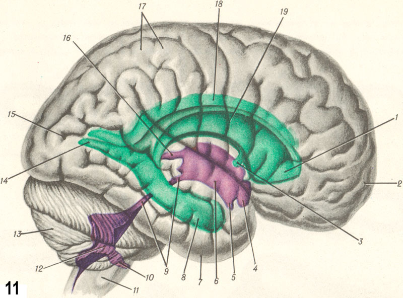
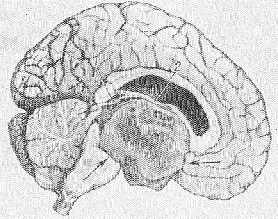
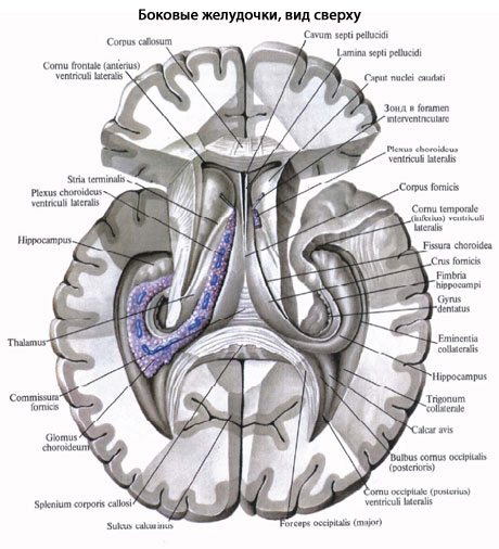
Lateral ventricles are located in the cerebral hemispheres (Fig. 2-4 and color. Fig. 11). They consist of a central part (pars centralis), the edges of which lie in the parietal lobe, and three processes extending from it on each side - horns. The anterior horn (cornu ant.) is located in the frontal lobe, the posterior horn (cornu post.) is in the occipital lobe, the lower horn (cornu inf.) is in the temporal lobe. The anterior horn has a triangular shape, bounded internally by a transparent septum (septum pellucidum), externally and posteriorly by the head of the caudate nucleus (caput nuclei caudati), and superiorly and anteriorly by the corpus callosum. Between the two plates of the transparent septum there is its cavity (cavum septi pellucidi). The central part of the ventricle has the shape of a slit, the bottom of the cut is formed by the caudate nucleus, the outer part of the upper surface of the thalamus and the terminal strip (stria terminalis) lying between them. Internally it is closed by an epithelial plate, covered from above by the corpus callosum. From the central part lateral ventricle the posterior horn extends posteriorly and the inferior horn extends downwards. The junction of the central part with the posterior and inferior horns is called the collateral triangle (trigonum collaterale). Hind horn lying among white matter the occipital lobe of the brain, has a triangular shape, gradually narrowing posteriorly; on its inner surface there are two longitudinal projections: the lower one is the bird's spur (calcar avis), corresponding to the calcarine groove, and the upper one is the bulb of the posterior horn (bulbus cornus post.), formed by the fibers of the corpus callosum. The inferior horn is directed downward and forward and ends at a distance of 10-14 mm from the temporal pole of the hemispheres. Its upper wall is formed by the tail of the caudate nucleus and the terminal strip. On the medial wall there is an elevation - the hippocampus (hippocampus), which is created due to the depression of the parahippocampal groove (gyrus parahippocampalis) lying deep from the surface of the hemisphere. The lower wall, or bottom of the horn, is limited by the white matter of the temporal lobe and bears a cushion - the collateral eminence (eminentia collateralis), corresponding to the outside of the collateral groove. From the medial side, the pia mater is invaginated into the lower horn, forming the choroid plexus of the lateral ventricle (plexus chorioideus ventriculi lat.). The lateral ventricles are closed on all sides, with the exception of the interventricular (Monroy's) foramen, through which the lateral ventricles are connected to the third ventricle and through it to each other.
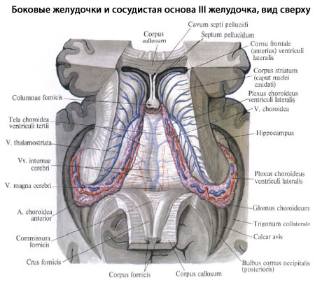
Third ventricle - unpaired cavity having a slit-like shape. Located in the diencephalon in the middle between the medial surfaces of the thalami and the hypothalamus. In front of the third ventricle there is the anterior commissure (commissura ant.), column of the fornix (columna fornicis), terminal plate (lamina terminalis); behind - posterior commissure (commissura post.), commissure of leashes (commissura habenularum); below - the posterior perforated substance (substantia perforata post.), gray tubercle (tuber cinereum), mastoid bodies (corpora mamillaria) and optic chiasm (chiasma opticum); on top is the vascular base of the third ventricle, attached to the upper surface of the thalamus, and above it are the crura fornicis, connected by the commissure of the fornix, and the corpus callosum. Lateral to the midline, the vascular base of the third ventricle contains the choroid plexus of the third ventricle (plexus chorioideus ventriculi tertii). In the middle of the third ventricle, the right and left thalamus are connected by an interthalamic fusion (adhesio interthalamica). The third ventricle forms depressions: the recess of the funnel (recessus infundibuli), the visual recess (recessus opticus), the epiphyseal recess (recessus pinealis). Through the aqueduct of the brain, the third ventricle is connected to the fourth.
Fourth ventricle. The bottom of the fourth ventricle, or rhomboid fossa (fossa rhomboidea), is formed by the cerebral pons (see) and the medulla oblongata (see), on the border of which the fourth ventricle forms lateral depressions (recessus lat. ventriculi quarti). The roof of the fourth ventricle (tegmen ventriculi quarti) has the shape of a tent and is composed of two cerebral sails - an unpaired upper one (velum medullare sup.), stretching between the upper cerebellar peduncles, and a paired lower one (velum medullare inf.), fixed to the legs of the flocculus (pedunculus flocculi) . Between the sails, the roof of the ventricle is formed by the cerebellum. The inferior medullary velum is covered with the vascular base of the fourth ventricle (tela chorioidea ventriculi quarti), with which the choroid plexus of the ventricle is connected. The cavity of the fourth ventricle communicates with the subarachnoid space by three openings: an unpaired median, located in the midline in the lower parts of the fourth ventricle, and paired lateral ones - in the area of the lateral recesses of the fourth ventricle. In the lower parts, the fourth ventricle, gradually narrowing, passes into the central canal of the spinal cord, which expands below into the terminal ventricle.
Pathology
Inflammatory processes in the gastrointestinal tract (ventriculitis) can be observed with various infectious lesions and intoxications of c. n. With. (for example, with meningoencephalitis, etc.). In acute ventriculitis, a picture of serous or purulent ependymatitis may develop (see Chorioependimitis). With hron, productive periventricular encephalitis, the ventricular ependyma thickens, sometimes taking on a granular appearance, which is caused by warty reactive growths of the subependymal layer. The course of ependymatitis is often aggravated due to disturbances in the circulation of cerebrospinal fluid due to obstruction of its outflow pathways at the level of the interventricular foramina, the cerebral aqueduct, and the unpaired median foramen of the fourth ventricle.
Clinically, disturbances in the circulation of cerebrospinal fluid in ventriculitis are manifested by paroxysms of headaches, during which patients, depending on the level of difficulty in the outflow of cerebrospinal fluid, take characteristic forced positions with the head tilted forward, tilted back, etc. (see Occlusive syndrome). Neurol, symptoms of ventriculitis are polymorphic; it manifests itself with a wide range of symptoms from the periventricular (periventricular) structures of the diencephalic parts of the brain (arterial hypertension, hyperthermia, diabetes insipidus, narcolepsy, cataplexy), midbrain (oculomotor disorders), hindbrain and medulla oblongata - the bottom of the fourth ventricle (vestibular disorders, symptoms of damage nuclei of VI,VII cranial nerves, etc.). In acute ventriculitis, cytosis is usually observed in the ventricular cerebrospinal fluid; in chronic ventriculitis, the ventricular fluid may be hydrocephalic (decreased protein content with a normal number of cells).
Primary hemorrhages in the gastrointestinal tract are rare and in the vast majority of cases are of traumatic origin. Secondary hemorrhages are more often observed, resulting from the breakthrough of intracerebral hematomas (traumatic, after a stroke) into the ventricular cavity. These hemorrhages are manifested by the acute development of a coma with pronounced reactions from the cardiovascular system, respiratory disorders, hyperthermia, dissociated meningeal symptoms, and often hormetonic syndrome (see Hormetonia). An admixture of blood is detected in the cerebrospinal fluid.


Brain, tumors). Tumors of the lateral ventricles are clinically manifested by a remitting course with occlusive-hydrocephalic paroxysms due to obstruction of the interventricular foramina. During paroxysms, a forced position of the head and symptoms of infringement of the brainstem are noted (upward gaze paralysis, bilateral pathological reflexes in the legs, disturbances in cardiovascular activity and breathing). Dissociated meningeal symptoms are often observed as a manifestation of tonic reflexes due to irritation of brain stem structures. In addition, periventricular symptoms may be detected as a result of the impact of the tumor on the adjacent parts of the brain (motor and sensory disturbances varying in severity over time, hemianopia, unilateral symptoms of subcortical lesions, general epileptic seizures with a tonic convulsive component, etc.). In the ventricular cerebrospinal fluid there is usually a sharp increase in protein, often combined with an increase in the number of cells and xanthochromia.
Tumors of the third ventricle are characterized by a combination of hypertensive-hydrocephalic symptoms due to occlusion of the cerebrospinal fluid circulation pathways - the cerebral aqueduct and interventricular (Monroy's) foramina with various metabolic-endocrine and autonomic-vascular disorders, which often serve as the first manifestations of the disease. Cataplectoid type seizures, sleep rhythm disturbances, sometimes patol, drowsiness are observed. In the later stages of the disease - attacks of decerebrate rigidity with respiratory and cardiovascular disorders. There is usually a significant increase in protein in the cerebrospinal fluid, sometimes with an increase in the number of cells and xanthochromia.
Wedge, the picture of a tumor of the fourth ventricle consists of symptoms of damage to the nuclear formations of the periventricular structures of its bottom and hypertensive-hydrocephalic symptoms due to obstruction of the outflow tract of cerebrospinal fluid. Paroxysms of headaches with vomiting, dizziness and impaired cardiovascular activity and breathing (Bruns' attacks) are characteristic. A constant symptom is pronounced brainstem nystagmus.
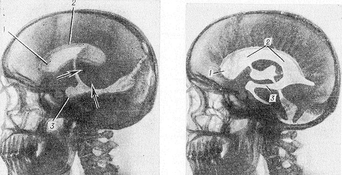
When diagnosing the pathology of gastrointestinal tract, in addition to analyzing the characteristics of the wedge, manifestations, ventriculography (see), ventriculoscopy (see) and encephalography (see) are used using water-soluble emulsion and gas radiopaque substances and radioisotopes (Fig. 8- 10).
Treatment
In inflammatory processes, surgical intervention is resorted to in cases of development of occlusive phenomena (see Hydrocephalus). Ventriculopuncture is used as a temporary measure for acute occlusions of the cerebrospinal fluid outflow tract to reduce intraventricular pressure (see).
In cases where the occlusion cannot be surgically eliminated, palliative operations are performed aimed at creating a roundabout path for the outflow of cerebrospinal fluid from the ventricles (ventriculostomy operations, perforation of the terminal plate, ventriculosubdural anastomosis, ventriculocisternostomy).
Among the conservative methods of treating ventriculitis, dehydration is used to reduce intracranial pressure and reduce hypertensive syndrome (see Dehydration therapy). For acute and chronic infectious ventriculitis, anti-inflammatory treatment is carried out.
Bibliography: Multi-volume guide to neurology, ed. S. N. Davidenkova, vol. 5, M., 1961; Multi-volume guide to surgery, ed. B.V. Petrovsky, vol. 3, book. 2, M., 1968; Patten B. M. Human embryology, trans. from English, M., 1959; Shelia R. N. Tumors of the ventricular system of the brain, L., 1973; G 1 a g a M. Das Nerven-system des Menschen, Lpz., 1953; G o r-rales M. a. T o r r e a 1 b a G. The third ventricle, Normal anatomy and changes in some pathological conditions, Neuroradiology, v. 11, p. 271, 1976, bibliogr.; Messert B., Wanna-maker B. B. a. Dudley A. W. Reevaluation of the size of the laterol ventricles of the brain, Postmortem study of an adult population, Neurology (Min-neap.), v. 22, p. 941, 1972.
E. P. Kononova, S. S. Mikhailov; N. Ya. Vasin (neurosurgeon).
In the hemispheres of the telencephalon, two lateral ventricles, ventriculi laterales, lie below the level of the corpus callosum, symmetrically on the sides of the midline, separated from the superolateral surface of the hemispheres by the entire thickness of the medulla.
The cavity of each lateral ventricle corresponds to the shape of the hemisphere: it begins in the frontal lobe in the form of an anterior horn, cornu anterius, curved downwards and to the lateral side, from here it stretches through the region of the parietal lobe under the name of the central part, pars centralis, which is divided at the level of the posterior edge of the corpus callosum on the lower horn, cornu inferius, (in the thickness of the temporal lobe) and the posterior horn, cornu posterius (in the occipital lobe).
The medial wall of the anterior horn is formed by the septum pellucidum, which separates the anterior horn from the same horn of the other hemisphere.
The lateral wall and partly the bottom of the anterior horn are occupied by the gray eminence, the head of the caudate nucleus, caput nuclei caudati, and the upper wall is formed by fibers of the corpus callosum. The roof of the central, narrowest part of the lateral ventricle also consists of fibers of the corpus callosum, while the bottom is made up of a continuation of the caudate nucleus, corpus nuclei caudati, and part of the upper surface of the thalamus.
The posterior horn is surrounded by a layer of white nerve fibers originating from the corpus callosum, the so-called tapetum; on its medial wall there is a noticeable ridge - a bird's spur, calcar avis, formed by depression from the side of the sulcus calcarinus, located on the medial surface of the hemisphere. The superolateral wall of the lower horn is formed by a tapetum, which is a continuation of the same formation surrounding the posterior horn. On the medial side on the upper wall there is a thinned part of the caudate nucleus, bent downwards and anteriorly - cauda nuclei caudati.

Along the medial wall of the lower horn it stretches along its entire length white the elevation is the hippocampus, hippocampus, which is formed as a result of depression from the sulcus hippocampi deeply embedded in the outside. The anterior end of the hippocampus is divided by grooves into several small tubercles. Along the medial edge of the hippocampus there is a so-called fimbria hippocampi, which is a continuation of the crus fornicis. At the bottom of the lower horn there is a ridge, eminentia collaterdlis, which comes from an indentation outside the groove of the same name.

From the medial side of the lateral ventricle, the pia mater protrudes into its central part and lower horn, forming in this place the choroid plexus, plexus choroideus ventriculi lateralis. The plexus is covered with epithelium, which represents the remnant of the undeveloped medial wall of the ventricle. Plexus choroideus ventriculi lateralis is the lateral margin of the body of choroidea ventriculi tertii.
Which doctors should I contact to examine the lateral ventricles of the brain:
Neurologist
Neurosurgeon
What diseases are associated with the lateral ventricles of the brain:
What tests and diagnostics need to be done for the lateral ventricles of the brain:
X-ray of the brain
Brain MRI
Dopplerography of cerebral vessels
Is something bothering you? Do you want to find out more detailed information about the lateral ventricles of the brain or do you need an examination? You can make an appointment with a doctor– clinic Eurolab always at your service! The best doctors They will examine you, advise you, provide the necessary assistance and make a diagnosis. You can also call a doctor at home. Clinic Eurolab open for you around the clock.
How to contact the clinic:
Phone number of our clinic in Kyiv: (+38 044) 206-20-00 (multi-channel). The clinic secretary will select a convenient day and time for you to visit the doctor. Our coordinates and directions are indicated. Look in more detail about all the clinic’s services on it.
If you have previously performed any research, Be sure to take their results to a doctor for consultation. If the studies have not been performed, we will do everything necessary in our clinic or with our colleagues in other clinics.
It is necessary to take a very careful approach to your overall health. There are many diseases that at first do not manifest themselves in our body, but in the end it turns out that, unfortunately, it is too late to treat them. To do this, you just need to do it several times a year. be examined by a doctor to not only prevent a terrible disease, but also maintain healthy mind in the body and the organism as a whole.
If you want to ask a doctor a question, use the online consultation section, perhaps you will find answers to your questions there and read self care tips. If you are interested in reviews about clinics and doctors, try to find the information you need on. Also register on the medical portal Eurolab to stay up to date latest news and updates of information about the Lateral Ventricles of the Brain on the website, which will be automatically sent to you by email.
Other anatomical terms starting with the letter "B":
| Thumb |
| Linea alba |
| Hip |
| Bronchi |
| Squirrels |
| Femur |
| Tibia |
| Brows |
| Eardrum |
| Labia majora |
The left (first) lateral ventricle is located in the left hemisphere, and the right (second) lateral ventricle is located in the right hemisphere of the cerebrum. The ventricular cavity has a complex shape. Its sections are located in all lobes of the hemisphere (with the exception of the insula). The parietal lobe of the cerebral hemisphere corresponds to the central part of the lateral ventricle, the frontal lobe - the anterior (frontal) horn, the occipital lobe - the posterior (occipital) horn, the temporal lobe - the lower (temporal) horn.
The central part, pars centralis, of the lateral ventricle is a horizontally located slit-like cavity, bounded above by transversely running fibers of the corpus callosum. The bottom of the central part is represented by the body of the caudate nucleus, part of the dorsal surface of the thalamus and the terminal stria, stria terminalis, separating the thalamus and caudate nucleus from each other.
The medial wall of the central part of the lateral ventricle is the body of the telencephalon. Between the body of the fornix above and the thalamus below there is a vascular fissure, fissura choroidea. Adjacent to the choroidal fissure from the central part is the choroid plexus of the lateral ventricle.
Laterally, the roof and the bottom of the central part of the lateral ventricle are connected at an acute angle. In this regard, the side wall of the central part is missing.
The anterior horn (frontal horn), cornu frontale (anterius), of the lateral ventricle has the appearance of a wide slit, curved downward and laterally. The medial wall of the anterior horn is the transparent septum. The lateral and partly lower walls of the anterior horn are formed by the head of the caudate nucleus. The anterior, superior and inferior walls of the anterior horn are bounded by fibers of the corpus callosum.
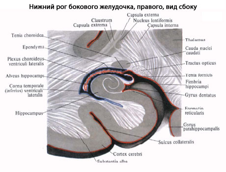
Lower horn ( temporal horn), cornu temporale (inferius), the lateral ventricle is the cavity of the temporal lobe. The lateral wall and roof of the inferior horn of the lateral ventricle are formed by the white matter of the cerebral hemisphere. The roof also includes the tail of the caudate nucleus, which continues here. In the area of the bottom of the lower horn, a collateral elevation, eminentia collateralis, continues from the posterior horn. This triangular-shaped elevation is a trace of the depression of parts of the cerebral hemisphere located in the depths of the collateral sulcus into the cavity of the lower horn. The medial wall of the inferior horn is formed by the hippocampus, hippocampus. The hippocampus extends to the most anterior parts of the inferior horn and ends in a thickening. This thickening of the hippocampus is divided by small grooves into separate tubercles (the toes of the seahorse - digitationes hippocampi (see fornix of the telencephalon, diagram, item 10). On the medial side, the hippocampus is fused with the hippocampus, fimbria hippocampi (see fornix of the telencephalon, diagram , item 6). The fimbria is a continuation of the pedicle of the fornix. The choroid plexus of the lateral ventricle descends here from the central part.
The posterior horn (occipital horn), cornu occipitale (posterius), of the lateral ventricle projects into the occipital lobe of the hemisphere. Its upper and lateral walls are formed by fibers of the corpus callosum, the lower and medial walls are formed by the protrusion of the white matter of the occipital lobe into the cavity of the posterior horn. Two protrusions are noticeable on the medial wall of the posterior horn. The upper one is the bulb of the posterior horn, bulbus cornu occipitdiis, represented by fibers of the corpus callosum on their way to the occipital lobe. The fibers of the corpus callosum in this place bend around the parieto-occipital groove, which protrudes into the depths of the hemisphere. The lower protrusion is the bird's spur, calcer avis, formed by pressing brain tissue into the cavity of the posterior horn from the depths of the calcarine groove. On the lower wall of the posterior horn there is a slightly convex collateral triangle, trigonum collaterale, a trace of the substance of the cerebral hemisphere located in the depths of the collateral sulcus being pressed into the ventricular cavity.
In the central part and lower horn of the lateral ventricle there is the choroid plexus of the lateral ventricle, plexus choroideus ventriculi lateralis. This plexus is attached to the vascular band, taenia choroidea, below and to the band of the fornix above. The choroid plexus continues into the inferior horn. Here it attaches to the fimbria of the hippocampus.
The choroid plexus of the lateral ventricle is formed due to the invagination into the ventricle through the vascular fissure, fissura choroidea, of the soft (choroid) membrane of the brain with the blood vessels contained in it. The soft membrane is covered on the side of the ventricle by the internal (epithelial) plate (the remnant of the medial wall of the first medullary bladder). In the anterior sections, the choroid plexus of the lateral ventricle through the interventricular foramen, foramen interventriculare, connects with the choroid plexus of the third ventricle.
|
Premise: |
Any
The human brain has several interconnected cavities filled with cerebrospinal fluid (CSF). These cavities are called ventricles. The ventricular system consists of two lateral ventricles connecting to the third ventricle, which, in turn, is connected through a thin canal (aqueduct of Sylvius) to the fourth ventricle. The fourth ventricle connects to the cavity of the spinal cord - the central canal, which is reduced in an adult.
Liquor is produced in the choroid plexuses of the ventricles and moves freely from the lateral ventricles to the fourth ventricle, and from there into the subarachnoid space of the brain and spinal cord, where it washes outer surface brain There it is reabsorbed into the bloodstream.
Lateral ventricles
The lateral ventricles are the cavities of the cerebral hemispheres (see Fig. 3.33). They are symmetrical slits in the thickness of the white matter containing cerebrospinal fluid. They have four parts corresponding to each lobe of the hemispheres: the central part - in the parietal lobe; anterior (frontal) horn - in the frontal lobe; posterior (occipital) horn – in the occipital lobe; the lower (temporal) horn is in the temporal lobe.
Central part looks like a horizontal slit. The upper wall (roof) of the central part is formed by the corpus callosum. At the bottom are the body of the caudate nucleus, partly the dorsal surface of the thalamus and the posterior leg of the fornix. In the central part of the lateral ventricles there is a developed choroid plexus of the lateral ventricle. It has the shape of a dark brown strip 4–5 mm wide. Posteriorly and downwardly it is directed into the cavity of the lower horn. The roof and bottom in the central part converge with each other at a very acute angle, i.e. There are no lateral walls near the central part of the lateral ventricles.
Front horn is a continuation of the central part and is directed forward and laterally. On the medial side it is limited by the plate of the septum pellucidum, on the lateral side by the head of the caudate nucleus. The remaining walls (anterior, superior and inferior) form the fibers of the forceps minor of the corpus callosum. The anterior horn has the widest lumen compared to other parts of the lateral ventricles.
Posterior horn has a pointed posterior shape with a convexity facing the lateral side. Its upper and lateral walls are formed by the fibers of the large forceps of the corpus callosum, and the remaining walls are represented by the white matter of the occipital lobe. There are two protrusions on the medial wall of the posterior horn: the upper one, called the dorsal horn bulb, corresponds to the parieto-occipital groove of the medial surface of the hemisphere, and the lower one, called the bird's spur, corresponds to the calcarine groove. The lower wall of the posterior horn has a triangular shape, slightly protruding into the cavity of the ventricle. Due to the fact that this triangular elevation corresponds to the collateral groove, it is called the “collateral triangle”.
Lower horn located in the temporal lobe and directed downward, forward and medially. Its lateral and superior walls are formed by the white matter of the temporal lobe of the hemisphere. The medial wall and partly the lower wall is occupied by the hippocampus. This elevation corresponds to the parahippocampal sulcus. A plate of white matter stretches along the medial edge of the hippocampus - the fimbria of the hippocampus, which is a continuation of the posterior leg of the fornix. On the lower wall (bottom) of the lower horn there is a collateral elevation, which is a continuation of the collateral triangle from the area of the posterior horn.
The lateral ventricles communicate with the third ventricle through the interventricular foramen (foramen of Monro). Through this opening, the choroid plexus penetrates from the cavity of the third ventricle into each lateral ventricle, which extends into the central part, the cavity of the posterior and inferior horns. The choroid plexuses of the ventricles of the brain produce cerebrospinal fluid. The shape and relationships of the ventricles of the brain are shown in Fig. 3.35.
Rice. 3.35.
a – lateral ventricles: 1 – anterior horn; 2 – corpus callosum; 3 – central part; 4 – posterior horn; 5 – lower horn; b – cast of the ventricular system of the brain: 1 – interventricular foramina; 2 – anterior horn; 3 – lower horn; 4 – third ventricle; 5 – cerebral aqueduct; 6 – fourth ventricle; 7 – posterior horn; 8 – central channel; 9 – median opening of the fourth ventricle; 10 – lateral openings of the fourth ventricle