In the hemispheres of the telencephalon, they lie below the level of the corpus callosum, symmetrically on the sides midline two lateral ventricles, ventriculi laterales, separated from the superolateral surface of the hemispheres by the entire thickness of the medulla.
Cavity of each lateral ventricle corresponds to the shape of the hemisphere: it begins in the frontal lobe in the form of an anterior horn, cornu anterius, curved downwards and to the lateral side, from here it stretches through the region of the parietal lobe under the name of the central part, pars centralis, which at the level of the posterior edge of the corpus callosum is divided into the inferior horn, cornu inferius, (in the thickness of the temporal lobe) and the posterior horn, cornu posterius (in the occipital lobe).
The medial wall of the anterior horn is formed by the septum pellucidum, which separates the anterior horn from the same horn of the other hemisphere.

The lateral wall and partly the bottom of the anterior horn are occupied by the gray eminence, the head of the caudate nucleus, caput nuclei caudati, and the upper wall is formed by fibers of the corpus callosum. The roof of the central, narrowest part of the lateral ventricle also consists of fibers of the corpus callosum, while the bottom is made up of the continuation of the caudate nucleus, corpus nuclei caudati, and part of the upper surface of the thalamus.

The posterior horn is surrounded by a layer of white nerve fibers originating from the corpus callosum, the so-called tapetum; on its medial wall there is a noticeable ridge - a bird's spur, calcar avis, formed by depression from the side of the sulcus calcarinus, located on the medial surface of the hemisphere. The superolateral wall of the lower horn is formed by a tapetum, which is a continuation of the same formation surrounding the posterior horn. On the medial side, on the upper wall, there is a thinned part of the caudate nucleus, bent downwards and anteriorly - cauda nuclei caudati.

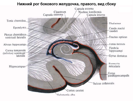
Along the medial wall of the lower horn it stretches along its entire length white the elevation is the hippocampus, hippocampus, which is formed as a result of depression from the sulcus hippocampi deeply embedded in the outside. The anterior end of the hippocampus is divided by grooves into several small tubercles. Along the medial edge of the hippocampus there is a so-called fimbria hippocampi, which is a continuation of the crus fornicis. At the bottom of the lower horn there is a ridge, eminentia collaterdlis, which comes from an indentation outside the groove of the same name.
From the medial side of the lateral ventricle, the pia mater protrudes into its central part and lower horn, forming in this place the choroid plexus, plexus choroideus ventriculi lateralis. The plexus is covered with epithelium, which represents the remnant of the undeveloped medial wall of the ventricle. Plexus choroideus ventriculi lateralis is the lateral margin of the body of choroidea ventriculi tertii.
Which doctors should I contact to examine the lateral ventricles of the brain:
Neurologist
Neurosurgeon
What diseases are associated with the lateral ventricles of the brain:
What tests and diagnostics need to be done for the lateral ventricles of the brain:
X-ray of the brain
Brain MRI
Dopplerography of cerebral vessels
Is something bothering you? Do you want to find out more detailed information about the lateral ventricles of the brain or do you need an examination? You can make an appointment with a doctor– clinic Eurolab always at your service! The best doctors They will examine you, advise you, provide the necessary assistance and make a diagnosis. You can also call a doctor at home. Clinic Eurolab open for you around the clock.
How to contact the clinic:
Phone number of our clinic in Kyiv: (+38 044) 206-20-00 (multi-channel). The clinic secretary will select a convenient day and time for you to visit the doctor. Our coordinates and directions are indicated. Look in more detail about all the clinic’s services on it.
If you have previously performed any research, Be sure to take their results to a doctor for consultation. If the studies have not been performed, we will do everything necessary in our clinic or with our colleagues in other clinics.
It is necessary to take a very careful approach to your overall health. There are many diseases that at first do not manifest themselves in our body, but in the end it turns out that, unfortunately, it is too late to treat them. To do this, you just need to do it several times a year. get examined by a doctor to not only prevent a terrible disease, but also maintain healthy mind in the body and the organism as a whole.
If you want to ask a doctor a question, use the online consultation section, perhaps you will find answers to your questions there and read self care tips. If you are interested in reviews about clinics and doctors, try to find the information you need on. Also register on the medical portal Eurolab to stay up to date latest news and updates of information about the Lateral Ventricles of the Brain on the website, which will be automatically sent to you by email.
Other anatomical terms starting with the letter "B":
| Thumb |
| Linea alba |
| Hip |
| Bronchi |
| Squirrels |
| Femur |
| Tibia |
| Brows |
| Eardrum |
| Labia majora |
In the hemispheres of the telencephalon, two lateral ventricles, ventriculi laterales, lie below the level of the corpus callosum, symmetrically on the sides of the midline, separated from the superolateral surface of the hemispheres by the entire thickness of the medulla.
The cavity of each lateral ventricle corresponds to the shape of the hemisphere: it begins in the frontal lobe in the form of an anterior horn, cornu anterius, curved downwards and to the lateral side, from here it stretches through the region of the parietal lobe under the name of the central part, pars centralis, which is divided at the level of the posterior edge of the corpus callosum on the lower horn, cornu inferius, (in the thickness of the temporal lobe) and the posterior horn, cornu posterius (in the occipital lobe).
The medial wall of the anterior horn is formed by the septum pellucidum, which separates the anterior horn from the same horn of the other hemisphere.
The lateral wall and partly the bottom of the anterior horn are occupied by the gray eminence, the head of the caudate nucleus, caput nuclei caudati, and the upper wall is formed by fibers of the corpus callosum. The roof of the central, narrowest part of the lateral ventricle also consists of fibers of the corpus callosum, while the bottom is made up of the continuation of the caudate nucleus, corpus nuclei caudati, and part of the upper surface of the thalamus.
The posterior horn is surrounded by a layer of white nerve fibers originating from the corpus callosum, the so-called tapetum; on its medial wall there is a noticeable ridge - a bird's spur, calcar avis, formed by depression from the side of the sulcus calcarinus, located on the medial surface of the hemisphere. The superolateral wall of the lower horn is formed by a tapetum, which is a continuation of the same formation surrounding the posterior horn. On the medial side, on the upper wall, there is a thinned part of the caudate nucleus, bent downwards and anteriorly - cauda nuclei caudati.
Along the medial wall of the lower horn, a white elevation stretches along its entire length - the hippocampus, hippocampus, which is formed as a result of depression from the sulcus hippocampi deeply embedded in the outside. The anterior end of the hippocampus is divided by grooves into several small tubercles. Along the medial edge of the hippocampus there is a so-called fimbria hippocampi, which is a continuation of the crus fornicis. At the bottom of the lower horn there is a ridge, eminentia collaterdlis, which comes from an indentation outside the groove of the same name.
From the medial side of the lateral ventricle, the pia mater protrudes into its central part and lower horn, forming in this place the choroid plexus, plexus choroideus ventriculi lateralis. The plexus is covered with epithelium, which represents the remnant of the undeveloped medial wall of the ventricle. Plexus choroideus ventriculi lateralis is the lateral margin of the body of choroidea ventriculi tertii.
Which doctors should I contact to examine the lateral ventricles of the brain:
Neurologist
Neurosurgeon
What diseases are associated with the lateral ventricles of the brain:
What tests and diagnostics need to be done for the lateral ventricles of the brain:
X-ray of the brain
Brain MRI
Dopplerography of cerebral vessels
Is something bothering you? Do you want to find out more detailed information about the lateral ventricles of the brain or do you need an examination? You can make an appointment with a doctor– clinic Eurolab always at your service! The best doctors will examine you, advise you, provide the necessary assistance and make a diagnosis. You can also call a doctor at home. Clinic Eurolab open for you around the clock.
How to contact the clinic:
Phone number of our clinic in Kyiv: (+38 044) 206-20-00 (multi-channel). The clinic secretary will select a convenient day and time for you to visit the doctor. Our coordinates and directions are indicated. Look in more detail about all the clinic’s services on it.
If you have previously performed any research, Be sure to take their results to a doctor for consultation. If the studies have not been performed, we will do everything necessary in our clinic or with our colleagues in other clinics.
It is necessary to take a very careful approach to your overall health. There are many diseases that at first do not manifest themselves in our body, but in the end it turns out that, unfortunately, it is too late to treat them. To do this, you just need to do it several times a year. get examined by a doctor, in order not only to prevent a terrible disease, but also to maintain a healthy spirit in the body and the organism as a whole.
If you want to ask a doctor a question, use the online consultation section, perhaps you will find answers to your questions there and read self care tips. If you are interested in reviews about clinics and doctors, try to find the information you need on. Also register on the medical portal Eurolab to keep abreast of the latest news and information updates about the Lateral Ventricles of the Brain on the website, which will be automatically sent to you by email.
Other anatomical terms starting with the letter "B":
| Thumb |
| Linea alba |
| Hip |
| Bronchi |
| Squirrels |
| Femur |
| Tibia |
| Brows |
| Eardrum |
| Labia majora |
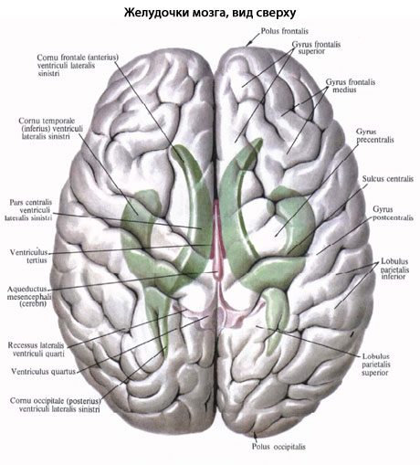
The left (first) lateral ventricle is located in the left hemisphere, and the right (second) lateral ventricle is located in the right hemisphere big brain. The ventricular cavity has a complex shape. Its divisions are located in all lobes of the hemisphere (with the exception of the insula). The parietal lobe of the cerebral hemisphere corresponds to the central part of the lateral ventricle, the frontal lobe - the anterior (frontal) horn, the occipital lobe - the posterior (occipital) horn, the temporal lobe - the lower (temporal) horn.
The central part, pars centralis, of the lateral ventricle is a horizontally located slit-like cavity, bounded above by transversely running fibers of the corpus callosum. The bottom of the central part is represented by the body of the caudate nucleus, part of the dorsal surface of the thalamus and the terminal strip, stria terminalis, separating the thalamus and caudate nucleus from each other.
The medial wall of the central part of the lateral ventricle is the body of the telencephalon. Between the body of the fornix above and the thalamus below there is a vascular fissure, fissura choroidea. Adjacent to the choroidal fissure from the central part is the choroid plexus of the lateral ventricle.
Laterally, the roof and the bottom of the central part of the lateral ventricle are connected under acute angle. In this regard, the side wall of the central part is missing.
The anterior horn (frontal horn), cornu frontale (anterius), of the lateral ventricle has the appearance of a wide slit, curved downward and laterally. The medial wall of the anterior horn is the transparent septum. The lateral and partly lower walls of the anterior horn are formed by the head of the caudate nucleus. The anterior, superior and inferior walls of the anterior horn are bounded by fibers of the corpus callosum.
The lower horn (temporal horn), cornu temporale (inferius), of the lateral ventricle is the cavity of the temporal lobe. The lateral wall and roof of the inferior horn of the lateral ventricle are formed by the white matter of the cerebral hemisphere. The roof also includes the tail of the caudate nucleus, which continues here. In the area of the bottom of the lower horn, a collateral elevation, eminentia collateralis, continues from the posterior horn. This triangular-shaped elevation is a trace of the depression of parts of the cerebral hemisphere located in the depths of the collateral sulcus into the cavity of the lower horn. The medial wall of the inferior horn is formed by the hippocampus, hippocampus. The hippocampus extends to the most anterior parts of the inferior horn and ends in a thickening. This thickening of the hippocampus is divided by fine grooves into individual tubercles (toes seahorse- digitationes hippocampi (see Fornix of the telencephalon, diagram, paragraph 10). On the medial side, the hippocampal fimbria, fimbria hippocampi, is fused with the hippocampus (see fornix of the telencephalon, diagram, paragraph 6). The fimbria is a continuation of the pedicle of the arch. Attached to the fimbria is the choroid plexus of the lateral ventricle, which descends here from the central part.
Posterior horn ( occipital horn), cornu occipitale (posterius), the lateral ventricle projects into the occipital lobe of the hemisphere. Its upper and lateral walls are formed by fibers of the corpus callosum, the lower and medial walls are formed by protrusion white matter occipital lobe into the cavity of the posterior horn. Two protrusions are noticeable on the medial wall of the posterior horn. The upper one is the bulb of the posterior horn, bulbus cornu occipitdiis, represented by fibers of the corpus callosum on their way to the occipital lobe. The fibers of the corpus callosum in this place bend around the parieto-occipital groove, which protrudes into the depths of the hemisphere. The lower protrusion is the bird's spur, calcer avis, formed by pressing brain tissue into the cavity of the posterior horn from the depths of the calcarine groove. On the lower wall of the posterior horn there is a slightly convex collateral triangle, trigonum collaterale, a trace of the substance of the cerebral hemisphere located in the depths of the collateral sulcus being pressed into the ventricular cavity.
In the central part and lower horn of the lateral ventricle there is the choroid plexus of the lateral ventricle, plexus choroideus ventriculi lateralis. This plexus is attached to the vascular band, taenia choroidea, below and to the band of the fornix above. The choroid plexus continues into the inferior horn. Here it attaches to the fimbria of the hippocampus.
The choroid plexus of the lateral ventricle is formed due to the invagination into the ventricle through the vascular fissure, fissura choroidea, of the soft (choroid) membrane of the brain with the blood vessels contained in it. The soft membrane is covered on the side of the ventricle by the internal (epithelial) plate (the remnant of the medial wall of the first medullary bladder). In the anterior sections, the choroid plexus of the lateral ventricle through the interventricular foramen, foramen interventriculare, connects with the choroid plexus of the third ventricle.
|
Premise: |
Any
The human brain has several interconnected cavities filled with cerebrospinal fluid (CSF). These cavities are called ventricles. The ventricular system consists of two lateral ventricles connecting to the third ventricle, which, in turn, is connected through a thin canal (aqueduct of Sylvius) to the fourth ventricle. The fourth ventricle connects to the cavity of the spinal cord - the central canal, which is reduced in an adult.
Liquor is produced in the choroid plexuses of the ventricles and moves freely from the lateral ventricles to the fourth ventricle, and from there into the subarachnoid space of the brain and spinal cord, where it washes outer surface brain There it is reabsorbed into the bloodstream.
Lateral ventricles
The lateral ventricles are the cavities of the cerebral hemispheres (see Fig. 3.33). They are symmetrical slits in the thickness of the white matter containing cerebrospinal fluid. They have four parts corresponding to each lobe of the hemispheres: the central part - in the parietal lobe; anterior (frontal) horn - in the frontal lobe; posterior (occipital) horn – in the occipital lobe; the lower (temporal) horn is in the temporal lobe.
Central part looks like a horizontal slit. The upper wall (roof) of the central part is formed by the corpus callosum. At the bottom are the body of the caudate nucleus, partially the dorsal surface of the thalamus and hind leg vault. In the central part of the lateral ventricles there is a developed choroid plexus of the lateral ventricle. It has the shape of a dark brown strip 4–5 mm wide. Posteriorly and downwardly it is directed into the cavity of the lower horn. The roof and bottom in the central part converge with each other at a very acute angle, i.e. There are no lateral walls near the central part of the lateral ventricles.
Front horn is a continuation of the central part and is directed forward and laterally. On the medial side it is limited by the plate of the septum pellucidum, on the lateral side by the head of the caudate nucleus. The remaining walls (anterior, superior and inferior) form the fibers of the forceps minor of the corpus callosum. The anterior horn has the widest lumen compared to other parts of the lateral ventricles.
Posterior horn has a pointed posterior shape with a convexity facing the lateral side. Its upper and lateral walls are formed by the fibers of the large forceps of the corpus callosum, and the remaining walls are represented by the white matter of the occipital lobe. There are two protrusions on the medial wall of the posterior horn: the upper one, called the dorsal horn bulb, corresponds to the parieto-occipital groove of the medial surface of the hemisphere, and the lower one, called the bird's spur, corresponds to the calcarine groove. The lower wall of the posterior horn has a triangular shape, slightly protruding into the cavity of the ventricle. Due to the fact that this triangular elevation corresponds to the collateral groove, it is called the “collateral triangle”.
Lower horn located in the temporal lobe and directed downward, forward and medially. Its lateral and superior walls are formed by the white matter of the temporal lobe of the hemisphere. The medial wall and partly the lower wall is occupied by the hippocampus. This elevation corresponds to the parahippocampal sulcus. Along the medial edge of the hippocampus stretches a plate of white matter - the hippocampal fimbria, which is a continuation of the posterior leg of the fornix. On the lower wall (bottom) of the lower horn there is a collateral elevation, which is a continuation of the collateral triangle from the area of the posterior horn.
The lateral ventricles communicate with the third ventricle through the interventricular foramen (foramen of Monro). Through this opening, the choroid plexus penetrates from the cavity of the third ventricle into each lateral ventricle, which extends into the central part, the cavity of the posterior and inferior horns. The choroid plexuses of the ventricles of the brain produce cerebrospinal fluid. The shape and relationships of the ventricles of the brain are shown in Fig. 3.35.
Rice. 3.35.
a – lateral ventricles: 1 – anterior horn; 2 – corpus callosum; 3 – central part; 4 – posterior horn; 5 – lower horn; b – cast of the ventricular system of the brain: 1 – interventricular foramina; 2 – anterior horn; 3 – lower horn; 4 – third ventricle; 5 – cerebral aqueduct; 6 – fourth ventricle; 7 – posterior horn; 8 – central channel; 9 – median opening of the fourth ventricle; 10 – lateral openings of the fourth ventricle