The activity of the body is a natural reflex reaction to a stimulus. Reflex– the body’s reaction to irritation of receptors, which is carried out with the participation of the central nervous system. The structural basis of the reflex is the reflex arc.
Reflex arc– series connected chain nerve cells, which ensures the implementation of a reaction, a response to irritation.
The reflex arc consists of six components: receptors, afferent (sensitive) path, reflex center, efferent (motor, secretory) path, effector (working organ), feedback.
Reflex arcs can be of two types:
1) simple - monosynaptic reflex arcs (reflex arc of the tendon reflex), consisting of 2 neurons (receptor (afferent) and effector), there is 1 synapse between them;
2) complex – polysynaptic reflex arcs. They consist of 3 neurons (there may be more) - a receptor, one or more intercalary and an effector.
The idea of a reflex arc as an expedient response of the body dictates the need to supplement the reflex arc with another link - a feedback loop. This component establishes a connection between the realized result of the reflex reaction and the nerve center that issues executive commands. With the help of this component, the open reflex arc closed.
Features of a simple monosynaptic reflex arc:
1) geographically close receptor and effector;
2) reflex arc two-neuron, monosynaptic;
3) nerve fibers of group Aα (70-120 m/s);
4) short time reflex;
5) muscles contracting according to the type of single muscle contraction.
Features of a complex monosynaptic reflex arc:
1) territorially separated receptor and effector;
2) three-neuron receptor arch (there may be more neurons);
3) the presence of nerve fibers of groups C and B;
4) muscle contraction according to the tetanus type.
Features of the autonomic reflex:
1) the interneuron is located in the lateral horns;
2) the preganglionic nerve pathway begins from the lateral horns, after the ganglion - the postganglionic;
3) the efferent path of the autonomic nervous arch reflex is interrupted by the autonomic ganglion, in which the efferent neuron lies.
The difference between the sympathetic nervous arch and the parasympathetic: the sympathetic nervous arch has a short preganglionic pathway, since the autonomic ganglion lies closer to the spinal cord, and the postganglionic pathway is long.
In the parasympathetic arc, the opposite is true: the preganglionic pathway is long, since the ganglion lies close to the organ or in the organ itself, and the postganglionic pathway is short.
When a reflex occurs, there is always a sequential spread of excitation from the formation of the perceptive action of the stimulus (from the receptor) towards the central nervous system (along centripetal paths) and then, after complex processes occurring within its limits, towards the central nervous system. nervous system(along centrifugal paths) to the working body (to the effector). 
An example of a reflex act
Using the example of the activity of the dog's salivary gland, one can study the path along which excitation spreads during the implementation of a reflex act. The corresponding research is carried out under conditions of vivisection (acute) experience.
The animal is immobilized in one way or another. A glass tube - a cannula - is inserted into the incision of the prepared gland duct. If the irritants do not act, then the gland is at rest and saliva is not released from the cannula. The experimenter immerses the tip of the animal's tongue in a weak acid solution. Saliva begins to flow from the cannula, indicating that the gland has become active.
Acid excites special sensory nerve endings located on the surface of the tongue, which perceive chemical influences. The resulting excitation along the centripetal fibers of the sensory nerve (n. lingualis) spreads along the central part of the reflex arc (in the medulla oblongata) and through the centrifugal fibers of the secretory nerve (chorda tympani) reaches the salivary gland. If the sensory nerve is cut, then immersing the tip of the tongue in the acid does not cause salivation, since the reflex arc will be interrupted at its centripetal link. If you start to get irritated electric shock the central end of the cut nerve, then the reflex secretion of saliva can again be caused.
After cutting the nerves leading to the salivary gland, i.e. after breaking the integrity of the arc in its centrifugal part, irritation of the centripetal nerve ceases to cause the effect. Irritation by current of the peripheral end of the cut central nerve, going directly to the gland, naturally causes salivation.
The formations that receive sites in the reflex reaction, in their entirety, constituting the directed path for reflex excitation, are defined by the concept of “reflex arc”. The individual links of the reflex arc are: receptor, effector (muscle or gland) and nerve cells with their processes.
Excitation that comes to the brain from any receptor along a complex system of pathways can go to any centrifugal path and reach any effector organ.
The central nervous system of animals and humans is characterized by a certain morphological and functional structure, thanks to which communication between any areas of the process is possible. All this is caused by the occurrence of naturally recurring reflex reactions that ensure the regulation of body functions. When we talk in the future about reflex muscular acts, about vascular reflexes, about respiratory reflexes, about reflex excitation of the glands of the digestive tract... We will have in mind the relationships developed in the process of evolution, in which the excitation that has arisen in certain parts of the body reaches certain areas of the central nervous system. From here, impulses are sent to certain organs and cause corresponding activity in them.
The course of excitation in the arc of the unconditioned reflex
We examined here the course of excitation in the arc, simplifying and schematizing the relationships and not taking into account the most complex processes that arise in the central part of the arc. In reality, a reflex act is almost never limited to a simple transfer of excitation from the centripetal part of the arc to the centrifugal one, as shown in the diagram. Excitation spreads much wider and involves various body systems in the reaction. For example, the entry of food substances into the mouth causes not only the secretory activity of the animal, on which we have focused our attention, but also motor activity, which captures a significant number of muscle effectors.
Conditioned reflex
Each excitation entering the central nervous system reaches its highest section, the cortex. cerebral hemispheres, and can become the basis for the formation of a temporary connection. In this case, we can talk about a friend of the conditioned reflex and build diagrams that reflect the fundamental side of the course of excitation during reflex activity of the cerebral cortex. However, consideration of such schemes should be attributed to the section of the course devoted to the special physiology of the cerebral hemispheres.
Here we only want to emphasize that no matter how complex the activity of the central nervous system is, we will always find in it elements characteristic of a simple reflex arc. This allows us to establish an evolutionary connection between the primitive nervous system of lower animals and the central nervous system of humans. The centripetal and centrifugal parts of the reflex arc retain fundamental similarities in the phylogenetic series of animals. In the process of evolution, predominantly the central part of the reflex pathway changed, which can be called the central nervous system in the narrowed sense of the word.
Briefly about the reflex arc
Basic form nervous activity is a reflex. Reflex is a causally determined reaction of the body to changes in the external or internal environment, carried out with the obligatory participation of the central nervous system in response to irritation of receptors. Due to reflexes, the emergence, change or cessation of any activity of the body occurs.
The neural pathway along which excitation spreads during reflexes is called reflex arc.
Reflex arcs consist of five components: 1) receptor; 2) afferent nerve pathway; 3) reflex center; 4) efferent nerve pathway; 5) effector (working body).
Receptor- This is a sensitive nerve ending that perceives irritation. In the receptors, the energy of the stimulus is converted into the energy of a nerve impulse. There are: 1) exteroceptors- are excited under the influence of stimuli from environment(receptors of the skin, eyes, inner ear, nasal mucosa and oral cavity); 2) interoreceptors- perceive irritations from the internal environment of the body (receptors internal organs, vessels); 3) proprioceptors- react to changes in position individual parts bodies in space (receptors of muscles, tendons, ligaments, joint capsules).
Afferent nerve pathway represented by processes of receptor neurons that carry excitations to the central nervous system.
Reflex center consists of a group of neurons located at various levels of the central nervous system and transmitting nerve impulses from the afferent to the efferent nerve pathway.
Efferent nerve pathway conducts nerve impulses from the central nervous system to the effector.
Effector- an executive organ whose activity changes under the influence of nerve impulses reaching it through the formations of the reflex arc. The effectors can be muscles or glands.
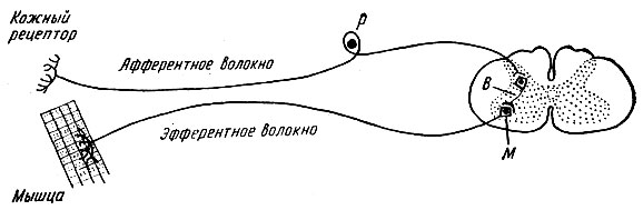
Reflex arcs can be simple or complex. A simple reflex arc consists of two neurons - a perceiver and an effector, between which there is one synapse. The diagram of such a two-neuron reflex arc is shown in Fig. 71.
An example of a simple reflex arc is the tendon reflex reflex arc, such as the knee reflex reflex arc.
The reflex arcs of most reflexes include not two, but a larger number of neurons: a receptor, one or more intercalary and an effector. Such reflex arcs are called complex, multineuron. The diagram of a complex (three-neuron) reflex arc is shown in Fig. 72.
It has now been established that during the response of the effector, numerous nerve endings present in the working organ are excited. Nerve impulses now from the effector again enter the central nervous system and inform it about the correctness of the response of the working organ. Thus, reflex arcs are not open, but circular formations.
Reflexes are very diverse. They can be classified according to a number of characteristics: 1) by biological significance(food, defensive, sexual); 2) depending on the type of receptors stimulated: exteroceptive, interoceptive and proprioceptive; 3) according to the nature of the response: motor or motor (executive organ - muscle), secretory (effector - gland), vasomotor (constriction or expansion of blood vessels).
All reflexes of the whole organism can be divided into two large groups: unconditioned and conditioned. The differences between them will be discussed in Chapter XII.
Functions of neurons. Classification of neurons.
Neuron (nerve cell)- basic structural and functional element nervous system; Humans have more than one hundred billion neurons. A neuron consists of a body and processes, usually one long process - an axon and several short branched processes - dendrites. Along dendrites, impulses follow to the cell body, along an axon - from the cell body to other neurons, muscles or glands. Thanks to the processes, neurons contact each other and form neural networks and circles through which nerve impulses circulate. A neuron, or nerve cell, is a functional unit of the nervous system. Neurons are susceptible to stimulation, that is, they are capable of being excited and transmitting electrical impulses from receptors to effectors. Based on the direction of impulse transmission, afferent neurons (sensory neurons), efferent neurons (motor neurons) and interneurons are distinguished. Each neuron consists of a soma (a cell with a diameter of 3 to 100 microns, containing a nucleus and other cellular organelles immersed in the cytoplasm) and processes - axons and dendrites. Based on the number and location of processes, neurons are divided into unipolar neurons, pseudounipolar neurons, bipolar neurons and multipolar neurons. .
The main functions of a nerve cell are the perception of external stimuli ( receptor function), their processing (integrative function) and transmission of nervous influences to other neurons or various working organs (effector function)
The peculiarities of the implementation of these functions make it possible to divide all neurons of the central nervous system into two large groups:
1) Cells that transmit information over long distances (from one part of the central nervous system to another, from the periphery to the center, from the center to executive body). These are large afferent and efferent neurons that have on their body and processes large number synapses, both inhibitory and excitatory, and capable of complex processes of processing influences coming through them.
2) Cells that provide interneural connections within organic nervous structures (intermediate neurons of the spinal cord, cerebral cortex, etc.). These are small cells that perceive nerve influences only through excitatory synapses. These cells are not capable of complex processes of integration of local synoptic influences of potentials; they serve as transmitters of excitatory or inhibitory influences on other nerve cells.
Perceiving function of a neuron. All irritations entering the nervous system are transmitted to the neuron through certain sections of its membrane located in the area of synaptic contacts. 6.2 Integrative function of a neuron. The overall change in the membrane potential of a neuron is the result of a complex interaction (integration) of local EPSPs and IPSPs of all the numerous activated synapses on the cell body and dendrites.
Effector function of a neuron. With the advent of AP, which, unlike local changes in membrane potential (EPSP and IPSP), is a spreading process, the nerve impulse begins to be conducted from the body of the nerve cell along the axon to another nerve cell or working organ, i.e. the effector function of the neuron is carried out.
Synapses in the central nervous system.
Synapse is a morphofunctional formation of the central nervous system, which ensures signal transmission from a neuron to another neuron or from a neuron to an effector cell. All CNS synapses can be classified as follows.
1. By localization: central and peripheral (neuromuscular, neurosecretory synapse of the autonomic nervous system).
2. According to development in ontogenesis: stable and dynamic, emerging in the process of individual development.
3. By final effect: inhibitory and excitatory.
4. According to the signal transmission mechanism: electrical, chemical, mixed.
5. Chemical synapses can be classified:
A) by contact form- terminal (flask-shaped connection) and transient (varicose dilatation of the axon);
b) by the nature of the mediator– cholinergic, adrenergic, dopaminergic
Electrical synapses. It is now recognized that there are electrical synapses in the central nervous system. From a morphological point of view, an electrical synapse is a gap-like formation (slit dimensions up to 2 nm) with ion bridges-channels between two contacting cells. Current loops, in particular in the presence of an action potential (AP), almost unhinderedly jump through such a gap-like contact and excite, i.e. induce the generation of APs in the second cell. In general, such synapses (they are called ephapses) provide very rapid transmission of excitation. But at the same time, with the help of these synapses it is impossible to ensure unilateral conduction, since most of these synapses have bilateral conductivity. In addition, they cannot be used to force an effector cell (a cell that is controlled through a given synapse) to inhibit its activity. An analogue of the electrical synapse in smooth muscles and in cardiac muscle are gap junctions of the nexus type.
Chemical synapses. In structure, chemical synapses are the ends of an axon (terminal synapses) or its varicose part (passing synapses), which is filled chemical- a mediator. In a synapse, there is a presynaptic element, which is limited by the presynaptic membrane, a postsynaptic element, which is limited by the postsynaptic membrane, as well as an extrasynaptic region and a synaptic cleft, the size of which is on average 50 nm.
Reflex arc. Classification of reflexes.
Reflex- the body’s reaction to changes in the external or internal environment, carried out through the central nervous system in response to irritation of receptors.
All reflex acts of the whole organism are divided into unconditioned and conditioned reflexes. Unconditioned reflexes are inherited, they are inherent in everyone biological species; their arches are formed at the time of birth and normally remain throughout life. However, they can change under the influence of illness. Conditioned reflexes arise with individual development and accumulation of new skills. The development of new temporary connections depends on changing environmental conditions. Conditioned reflexes are formed on the basis of unconditioned ones and with the participation of higher parts of the brain. They can be classified into various groups according to a number of signs.
1. According to biological significance
A.) food
B.) defensive
B.) sexual
G.) approximate
D.) postural-tonic (reflexes of body position in space)
E.) locomotor (reflexes of body movement in space)
2. By location of receptors, the irritation of which is caused by this reflex act
A.) exteroceptive reflex - irritation of receptors on the external surface of the body
B.) viscero- or interoreceptive reflex - arising from irritation of receptors of internal organs and blood vessels
B.) proprioceptive (myotatic) reflex - irritation of receptors skeletal muscles, joints, tendons
3. According to the location of the neurons involved in the reflex
A.) spinal reflexes - neurons located in the spinal cord
B.) bulbar reflexes - carried out with the obligatory participation of neurons of the medulla oblongata
B.) mesencephalic reflexes - carried out with the participation of midbrain neurons
D.) diencephalic reflexes - neurons of the diencephalon are involved
D.) cortical reflexes - carried out with the participation of neurons in the cerebral cortex
Reflex arc- this is the path along which irritation (signal) from the receptor passes to the executive organ. The structural basis of the reflex arc is formed by neural circuits consisting of receptor, intercalary and effector neurons. It is these neurons and their processes that form the path along which nerve impulses from the receptor are transmitted to the executive organ during the implementation of any reflex.
In the peripheral nervous system, reflex arcs (neural circuits) are distinguished
Somatic nervous system, innervating the skeletal muscles
The autonomic nervous system innervates internal organs: heart, stomach, intestines, kidneys, liver, etc.
The reflex arc consists of five sections:
1. Receptors that perceive irritation and respond to it with excitement. Receptors are located in the skin, in all internal organs; clusters of receptors form the sense organs (eye, ear, etc.).
2. Sensitive (centripetal, afferent) nerve fiber, transmitting excitation to the center; a neuron that has this fiber is also called sensitive. The cell bodies of sensory neurons are located outside the central nervous system - in ganglia along the spinal cord and near the brain.
3. Nerve center, where excitation switches from sensory neurons to motor neurons; The centers of most motor reflexes are located in the spinal cord. The brain contains centers for complex reflexes, such as protective, food, orientation, etc. In the nerve center
There is a synaptic connection between the sensory and motor neurons.
1. Motor (centrifugal, efferent) nerve fiber, carrying excitation from the central nervous system to the working organ; Centrifugal fiber is a long extension of a motor neuron. A motor neuron is a neuron whose process approaches the working organ and transmits a signal to it from the center.
2. Effector - a working organ that produces an effect, a reaction in response to receptor irritation. Effectors can be muscles that contract when they receive stimulation from the center, gland cells that secrete juice under the influence of nervous stimulation, or other organs.
The concept of the nerve center.
Nerve center- a set of nerve cells, more or less strictly localized in the nervous system and certainly involved in the implementation of a reflex, in the regulation of one or another function of the body or one of the aspects of this function. In the simplest cases, the nerve center consists of several neurons forming a separate node (ganglion).
In every N. c. Through the input channels - the corresponding nerve fibers - information from the sense organs or from other nervous systems arrives in the form of nerve impulses. This information is processed by the neurons of the central nervous system, whose processes (axons) do not extend beyond its boundaries. The final link is the neurons, the processes of which leave the N. c. and deliver its command impulses to peripheral organs or other N. c. (output channels). The neurons that make up the neural network are connected to each other through excitatory and inhibitory synapses and form complex complexes, so-called neural networks. Along with neurons that are excited only in response to incoming nerve signals or the action of various chemical stimuli contained in the blood, the composition of N. c. may include pacemaker neurons that have their own automaticity; They have the ability to periodically generate nerve impulses.
Localization of N. c. determined on the basis of experiments with irritation, limited destruction, removal or transection of certain parts of the brain or spinal cord. If, when a given area of the central nervous system is irritated, one or another physiological reaction occurs, and when it is removed or destroyed, it disappears, then it is generally accepted that the central nervous system is located here, influencing this function or participating in a certain reflex.
Properties of nerve centers.
The nerve center (NC) is a collection of neurons in various parts of the central nervous system that provide regulation of any function of the body.
The following features are characteristic for conducting excitation through nerve centers:
1. Single-line conduction, it goes from the afferent, through the intercalary to the efferent neuron. This is due to the presence of interneuron synapses.
2. The central delay in the conduction of excitation, i.e., along the NC excitation is much slower than along the nerve fiber. This is explained by synaptic delay because most of the synapses are in the central link of the reflex arc, where the conduction speed is the lowest. Based on this, reflex time is the time from the onset of exposure to the stimulus to the appearance of the response. The longer the central delay, the more time reflex. However, it depends on the strength of the stimulus. The larger it is, the shorter the reflex time and vice versa. This is explained by the phenomenon of summation of excitations in synapses. In addition, it is determined by the functional state of the central nervous system. For example, when the NC is tired, the duration of the reflex reaction increases.
3. Spatial and temporal summation. Temporal summation occurs, as in synapses, due to the fact that the more nerve impulses arrive, the more neurotransmitter is released in them, the higher the EPSP amplitude. Therefore, a reflex reaction can occur to several successive subthreshold stimuli. Spatial summation is observed when impulses from several neuron receptors go to the nerve center. When subthreshold stimuli act on them, the resulting postsynaptic potentials are summed up 11 and a propagating AP is generated in the neuron membrane.
4. Transformation of the rhythm of excitation - a change in the frequency of nerve impulses when passing through the nerve center. The frequency may decrease or increase. For example, increasing transformation (increase in frequency) is due to the dispersion and multiplication of excitation in neurons. The first phenomenon occurs as a result of the division of nerve impulses into several neurons, the axons of which then form synapses on one neuron. Second, the generation of several nerve impulses during the development of an excitatory postsynaptic potential on the membrane of one neuron. The downward transformation is explained by the summation of several EPSPs and the appearance of one AP in the neuron.
5. Post-tetanic potentiation, this is an increase in the reflex response as a result of prolonged excitation
center neurons. Under the influence of many series of nerve impulses passing at high frequency through the synapses, a large amount of neurotransmitter is released at the interneuron synapses. This leads to a progressive increase in the amplitude of the excitatory postsynaptic potential and long-term (several hours) excitation of neurons.
6. Aftereffect is a delay in the end of the reflex response after the cessation of the stimulus. Associated with the circulation of nerve impulses along closed circuits of neurons.
7. The tone of the nerve centers is a state of constant increased activity. It is caused by the constant supply of nerve impulses to the NC from peripheral receptors, the stimulating influence of metabolic products and other humoral factors on neurons. For example, the manifestation of the tone of the corresponding centers is the tone of a certain muscle group.
8. automaticity or spontaneous activity of nerve centers. Periodic or constant generation of nerve PULSES by neurons, which arise spontaneously in them, i.e. in the absence of signals from other neurons or receptors. It is caused by fluctuations in the metabolic processor in neurons and the effect of humoral factors on them.
9. Plasticity of nerve centers. This is their ability to change functional properties. In this case, the center acquires the ability to perform new functions or restore old ones after damage. The basis of plasticity N.Ts. lies the plasticity of synapses and membranes of neurons, which can change their molecular structure.
10. Low physiological lability and fatigue. N.Ts. can conduct pulses of only a limited frequency. Their fatigue is explained by fatigue of synapses and deterioration of neuronal metabolism.
Inhibition in the central nervous system.
Inhibition in the central nervous system prevents the development of excitation or weakens ongoing excitation. An example of inhibition could be the cessation of a reflex reaction against the background of the action of another stronger stimulus. Initially, a unitary-chemical theory of inhibition was proposed. It was based on Dale's principle: one neuron - one transmitter. According to it, inhibition is provided by the same neurons and synapses as excitation. Subsequently, the correctness of the binary chemical theory was proven. According to the latter, inhibition is provided by special inhibitory neurons, which are intercalary. These are Renshaw cells of the spinal cord and Purkinje neurons. Inhibition in the central nervous system is necessary for the integration of neurons into a single nerve center. The following inhibitory mechanisms are distinguished in the central nervous system:
1| Postsynaptic. It occurs in the postsynaptic membrane of the soma and dendrites of neurons, i.e. after the transmitting synapse. In these areas, specialized inhibitory neurons form axo-dendritic or axosomatic synapses (Fig.). These synapses are glycinergic. As a result of the effect of NLI on the glycine chemoreceptors of the postsynaptic membrane, its potassium and chloride channels open. Potassium and chloride ions enter the neuron, and IPSP develops. The role of chlorine ions in the development of IPSP: small. As a result of the resulting hyperpolarization, the excitability of the neuron decreases. The conduction of nerve impulses through it stops. The alkaloid strychnine can bind to glycerol receptors on the postsynaptic membrane and turn off inhibitory synapses. This is used to demonstrate the role of inhibition. After the administration of strychnine, the animal develops cramps in all muscles.
2. Presynaptic inhibition. In this case, the inhibitory neuron forms a synapse on the axon of the neuron that approaches the transmitting synapse. Those. such a synapse is axo-axonal (Fig.). The mediator of these synapses is GABA. Under the influence of GABA, chloride channels of the postsynaptic membrane are activated. But in this case, chlorine ions begin to leave the axon. This leads to a small local but long-lasting depolarization of its membrane.
A significant part of the sodium channels of the membrane is inactivated, which blocks the conduction of nerve impulses along the axon, and consequently the release of the neurotransmitter at the transmitting synapse. The closer the inhibitory synapse is located to the axon hillock, the stronger its inhibitory effect. Presynaptic inhibition is most effective in information processing, since the conduction of excitation is not blocked in the entire neuron, but only at its one input. Other synapses located on the neuron continue to function.
3. Pessimal inhibition. Discovered by N.E. Vvedensky. Occurs at a very high frequency of nerve impulses. A persistent, long-term depolarization of the entire neuron membrane and inactivation of its sodium channels develops. The neuron becomes unexcitable.
Both inhibitory and excitatory postsynaptic potentials can simultaneously arise in a neuron. Due to this, the necessary signals are isolated.
The reflex arc consists of:
– receptors that perceive irritation.
– sensitive (centripetal, afferent) nerve fiber transmitting excitation to the center
– the nerve center where excitation switches from sensory neurons to motor neurons
– motor (centrifugal, efferent) nerve fiber, carrying excitation from the central nervous system to the working organ
– effector - a working organ that carries out an effect, a reaction in response to stimulation of the receptor.
Receptors and receptive fields
Receptor- cells that perceive irritation.
Receptive field- this is the anatomical area, when irritated, this reflex is caused.
The receptive fields of the primary sensory receptors are organized most simply. For example, the tactile or nociceptive receptive field of the skin surface represents the branches of a single sensory fiber.
Receptors located in different parts of the receptive field have different sensitivity to adequate stimulation. In the center of the receptive field there is usually a highly sensitive zone, and closer to the periphery of the receptive field sensitivity decreases.
The receptive fields of secondary sensory receptors are organized in a similar way. The difference is that the branches of the afferent fiber do not end freely, but have synaptic contacts with sensitive cells - receptors. This is how gustatory, vestibular, and acoustic receptive fields are organized.
Overlap of receptive fields. The same area of the sensitive surface (for example, the skin or retina) is innervated by several sensory nerve fibers, which with their branches overlap the receptive fields of individual afferent nerves.
By overlapping receptive fields, the total sensory surface of the body increases.
Classification of reflexes.
By type of education:
Conditional (acquired) - respond to the name, the dog saliva into the light.
Unconditional (congenital) - blinking, swallowing, knee.
By location receptors:
Exteroceptive (cutaneous, visual, auditory, olfactory),
Interoceptive (from receptors of internal organs)
Proprioceptive (from receptors of muscles, tendons, joints)
By effector:
Somatic, or motor, (skeletal muscle reflexes);
Autonomic internal organs - digestive, cardiovascular, excretory, secretory, etc.
By biological origin:
Defensive, or defensive (response to tactile pain)
Digestive (irritation of oral cavity receptors.)
Sexual (hormones in the blood)
Approximate (rotation of head, body)
Motor
Posotonic (supported body postures)
By the number of synapses:
Monosynaptic, the arcs of which consist of afferent and efferent neurons (for example, knee).
Polysynaptic, the arcs of which also contain 1 or more intermediate neurons and have 2 or more synaptic switches. (somat. and veget. refs).
Disynaptic (2 synapses, 3 neurons).
By the nature of the response:
Motor\motor (muscle contractions)
Secretory (secretory gland)
Vasomotor (dilation and constriction of blood vessels)
Cardiac (measures the work of the heart muscle.)
By duration:
phasic (fast) hand withdrawal
tonic (slow) posture maintenance
According to the location of the nerve center:
Spinal (SM neurons are involved) - withdrawal of the Hands from hot segments 2-4, knee reflex.
Reflexes in the brain
Bulbar (medulla oblongata) - closing of the eyelids when touching. to the cornea.
Mesencial (middle m) - vision landmark.
Diencephalic (diencephalon) – sense of smell
Cortical (BP GM cortex) – conditional. ref.
Properties of nerve centers.
1. One-sided propagation of excitation.
Excitation is transmitted from afferent to efferent neuron (reason: structure of the synapse).
Slowing down the transfer of excitation.
Conditional The presence of many synapses also depends on the strength of the stimulation (summation) and on the physical state. CNS (fatigue).
3.Summation addition of effects below threshold stimuli.
Temporary: ref. From prev. The imp-sa has not yet passed, but the trail. Already arrived.
Spatial: mixing several. Backup They are conditioned. Images Ref.
Center of relief and occlusion.
Center relief - occurs under the action of an optimal stimulus (max response) - appears. Relief center.
When the action is min. (lower response. Rection) occlusion has occurred.
Assimilation and transformation of the rhythm of excitation.
Transformation is a change in the frequency of a nerve impulse as it passes through the nerve center. The frequency can go up or down.
Assimilation (dance, daily routine)
Consequence
Delay in the end of a response after the cessation of stimulation. Associated with the circular nerve. Imp. By closed Circuits of neurons.
Short-term (fractions of a second)
long (seconds)
Rhythmic activity of nerve centers.
An increase or decrease in the frequency of nerve impulses associated with the properties of the synapse and the integrative duration of neurons.
8. Plasticity of nerve centers.
The ability to rebuild the functionality of a property for more effective regulation of functions, the implementation of new reflexes that were not previously characteristic of this center or the restoration of functional functions. The layer of synpses is based on a change in the malleolus structure.
Changes in excitability under the influence of chemicals.
High sensitivity to the effects of different things.
Fatigue of the nerve centers.
Associated with high synaptic fatigue. Decreased feelings. Receptors.
General principles coordination activities of the central nervous system.
Braking- special ner. proc. manifested itself in a decrease or complete disappearance of the response. reactions.
The principle of convergence
Convergence is the convergence of impulses arriving along various afferent pathways in any one central neuron or nerve center.
2 . The principle of convergence is closely related to the principle common final path open Sherrinkton. Many different stimuli can excite the same motor neuron and cause the same motor response. This principle is due to the unequal number of afferent and efferent pathways.
Divergence principle
This is the contact of one neuron with many others.
Irradiation and concentration of excitation.
The spread of the excitation process to other nerve centers is called irradiation (electoral- in one direction , generalized- extensive).
The irradiation after some time is replaced by the phenomenon of concentration of excitation in the same source point of the central nervous system.
The process of irradiation plays a positive role (formation of new conditioned reflexes) and negative (violation of the subtle relationships that have developed between the processes of excitation and inhibition, which leads to a disorder of motor activity) roles.
The principle of reciprocity (inhibits)
Excitation of some cells causes inhibition of others through an interneuron.
The principle of dominance
Ukhtomsky formulated the principle of dominance as a working principle of the activity of nerve centers. The term dominant denotes the dominant focus of excitation of the central nervous system, which determines the current activity of the body.
Principles of dominant focus :
Increased excitability of nerve centers;
Persistence of excitation over time;
Ability to summarize extraneous stimuli;
Inertia (the ability to maintain arousal for a long time after the end of the stimulus); the ability to cause conjugate inhibition.