The simplest reaction of the nervous system is reflex. It is a quick, automatic, stereotypical reaction to irritation, it is called an involuntary act, since it is not under the control of consciousness. Neurons that form the path of nerve impulses during a reflex act make up reflex arc. The simplest reflex arc in animals includes one neuron and has the following form:
Neuron Stimulus → Receptor - Effector → Response
This level of organization is characteristic of the nervous system of coelenterates. Reflex arcs of all groups of animals with a higher level of structural and functional organization consist of at least two neurons - afferent, or sensory(sensitive), conducting impulses from the receptor, and efferent, or motor(motor), transmitting impulses to the effector. Between these two neurons there may also be interneurons located in a cluster of nerve cells - a ganglion, a nerve chain, or the central nervous system (Fig. 16.13). There are a huge variety of reflexes of varying structural and functional complexity, but they can all be divided into the following four groups:
1. Monosynaptic reflexes. These are reflexes with the simplest arc found in vertebrates. The sensory neuron is in direct contact with the body of the motor neuron. Only one synapse, located in the central nervous system, participates in such an arc. Such reflexes are quite common in all vertebrates; they are involved in the regulation of muscle tone and posture (for example, the knee reflex - leg extension in knee joint). In these reflex arcs, neurons do not reach the brain, and reflex acts are carried out without its participation, since they are stereotypical and do not require thinking or conscious decision. They are economical in the number of central neurons involved and dispense with the intervention of the brain, which can “focus” on more important matters.
2. Polysynaptic spinal reflexes. At least two synapses located in the central nervous system participate in such reflexes, since a third neuron is included in the arc - intercalary, or intermediate(interneuron). Synapses are present here between sensory and interneuron neurons and between intercalary and motor neurons (Fig. 16.13, B). This type of reflex act serves as an example of a simple reflex that closes in the spinal cord. In Fig. Figure 16.14 presents in a highly simplified form the reflex that occurs when a finger is pricked with a pin.

Simple reflex arcs types 1 and 2 allow the body to carry out the automatic involuntary responses necessary to adapt to change external environment(for example, the pupillary reflex or maintaining balance when moving) and to changes in the body itself (regulation of respiratory rate, blood pressure, etc.), as well as to prevent damage to the body, such as injury or burn.
3. Polysynantic reflexes involving both the spinal cord and the brain. In this type of reflex arc, a sensory neuron forms a synapse in the spinal cord with a second neuron that sends impulses to the brain. Thus, these second sensory neurons form the ascending nerve pathways (Fig. 16.15A). The brain interprets this sensory information and stores it for later use. Along with this, he is at any time at the moment can initiate motor activity, and then the impulses will be transmitted by the motor neurons along the descending nerve pathway directly to the spinal motor neurons through synapses located in the same area as the output synapses of the interneurons (Fig. 16.15).
![]()
4. Conditioned reflexes. Conditioned reflexes are a type of reflex activity in which the nature of the response depends on past experience. These reflexes are coordinated by the brain. The basis of all conditioned reflexes (such as the habit of toileting, salivation at the sight and smell of food, awareness of danger) is learning (section 16.9).
There are many situations where one of two possible reflex reactions occurs, involving a particular group of muscles that can either contract or relax, which would lead to opposite results. In this situation, the usual spinal reflex would be carried out by the reflex arc shown in Fig. 16.14, however, the “conditions” under which the stimulus operates can change the response. In such cases, a more complex reflex arc operates, including both excitatory and inhibitory neurons. For example, if we grab with our hand an empty metal frying pan that turns out to be too hot and burns our fingers, we will probably immediately let it go, but we will carefully and quickly put the equally hot food on an expensive dish that burns our fingers in its place. The difference in response indicates that we are dealing with conditioned reflex, which involves memory and a conscious decision made by the brain. In this situation, the response is carried out along a more complex reflex pathway, shown in Fig. 16.16.

In both cases, the stimulus causes impulses that travel to the sensory part of the brain along the ascending nerve pathway. When these impulses enter the brain, it analyzes them, taking into account information coming from other senses, such as the eyes, and determines reason stimulus. The information entering the brain is compared with that which is already stored in it - with information about what will most likely happen if the spinal reflex is carried out automatically. In the case of a metal frying pan, the brain will calculate that if it is thrown, it will not cause any harm to the body or the frying pan, and will send impulses along excitatory pathway. This path goes down the spinal cord to the level where the stimulus entered the spinal cord and forms connections with the bodies of the motor neurons that carry out this reflex. The speed of impulses along this path is such that impulses from the excitatory motor neuron of the brain reach a special motor neuron simultaneously with impulses from the interneuron of a simple reflex arc. The effects of these and other impulses are summed up, and exciting impulses are sent to the muscle effector along the axon of the spinal motor neuron, causing them to throw the frying pan.
But in the case of a hot dish, the brain will quickly figure out that if you throw it, you can scald your legs, and besides, the food will be spoiled and the expensive dish will be broken. If you hold the dish and carefully place it in place, this will not cause severe burns to your fingers. After the brain makes such a decision, impulses will arise in it, which will also be transmitted to the spinal motor neurons, but this time along the inhibitory pathway. They will arrive simultaneously with excitatory impulses from the interneuron and extinguish their action. As a result, no impulses will flow through the motor neurons to the corresponding muscles and the dish will be held in the hands. At the same time, the brain can give the muscles a different program of action, and the dish will be quickly and carefully put in place.
The above description of reflex arcs is naturally greatly simplified. After all, the process of coordination, integration and regulation of functions in the body is much more complex. So, for example, certain neurons connect different levels of the spinal cord that control, say, the arms and legs, so that the activity of one level is coordinated with the activity of another, and another group of neurons exercises overall control from the brain.
While joint activities brain and endocrine system plays an important role in coordinating many types of nervous activity, described later in this chapter, the regulation of autonomic functions is carried out by another reflex system, which is based exclusively nervous activity. This system is called the autonomic or autonomic nervous system.
ESSENCE AND IMPORTANCE OF THE PROCESS OF SELF-REGULATION
FOR SELF-SATISFACTION OF HUMAN NEEDS
Homeostasis- the ability of biological systems to resist changes and maintain dynamic equilibrium internal environment of the body. The functional activity of organs and the coordination of their activities is supported by the nervous and (or) humoral regulatory systems. Nervous regulation carried out using nerve impulses and ensures a quick response of a certain organ to irritation. Humoral regulation carried out through the production of endocrine glands of biologically active substances (hormones, etc.) that enter the blood and affect the speed and direction of metabolic processes.
PC. Anokhin believed: function- is the achievement by an organism of a useful adaptive result in interaction with its environment. Functional system- such organization of activities individual parts organism, which ultimately produces a useful adaptive result.
Having created a behavior program, the body fights for it, overcoming the resistance of the environment, the features of which are taken into account through sensory amendments made to the program. As a result, behavior is actively restructured according to the principle feedback. Physiological basis of feedback - reflex ring. The main purpose of feedback is to minimize (reach a minimum or bring to it) any deviation of the controlled indicator from the norm. This is how regulatory systems maintain manageable indicators at a constant level.
Highest level regulation physiological functions and interaction of the body with the external environment are provided by the central nervous system. The ANS, controlling the functions of organs and systems, suppressing them or stimulating them, carries out II level of regulation. The endocrine system makes up III level of regulation, influencing regulatory processes with the help of hormones and other biologically active substances. Lower levels internal management provide automatic regulation systems (self-regulation), which support a certain mode of life using general physical and chemical laws. Interneuronal and neuromuscular intercellular contacts provide mediators.
Knowledge of the regulatory mechanisms of life is important for understanding the characteristics of the body’s adaptation to changing environmental conditions.
GENERAL PLAN OF THE STRUCTURE OF THE NERVOUS SYSTEM
The nervous system plays an important role in regulating the functions of the body and integrating the activities of its organs and systems. It communicates the body with the external environment. Studying the structure and functions of the nervous system is necessary to understand the mechanisms of development of human diseases and the proper organization of work and rest.
The nervous system consists of central And peripheral parts. Central nervous system (CNS) presented brain localized in the skull, and spinal cord located in the spinal canal. The brain and spinal cord are composed of white and gray matter. Gray matter represented by neurons and their dendrites. White matter consists of processes of nerve cells - nerve fibers (their white caused by myelin sheaths).
Nerve fibers form pathways connecting various parts of the central nervous system, nuclei (nerve centers) with each other.
Peripheral nervous system includes: spinal nerve roots; spinal and cranial nerves, their branches, nerve ganglia and plexuses.
Depending on the zones of innervation and some anatomical and physiological features, somatic and vegetative nervous system. Somatic nervous system provides innervation of the body - soma: skin and skeletal muscles. It regulates the body’s connections with the external environment using the senses.
Autonomic nervous system (ANS) innervates internal organs and glands, regulating metabolic processes in all tissues and organs, penetrating into them through the vessels. In the structure of the ANS there are sympathetic And parasympathetic departments, and in them, in turn, central and peripheral.
REFLEX. REFLECTOR ARC
The basis of nervous regulation is reflex activity. Reflex- the body’s response to irritation, carried out with the obligatory participation of the nervous system. The reflex response, in turn, acts on the stimulus. Thus, a reflex is a feedback process.
Reflex arc - the path of excitation from the receptor to the working organ - includes the receptor, the afferent nerve pathway, the reflex center, the efferent nerve pathway, and the effector. Impulses arise in receptors- sensitive nerve endings. Along the afferent (centripetal) path, nerve impulses from the receptors enter the reflex nerve center of the central nervous system. Afferent pathway represented by sensory nerve fibers of the afferent neuron. IN reflex center impulses are processed and switched to the efferent pathway. By efferent (centrifugal) pathway motor (effector) impulses from the nerve center reach the executive organ on the muscle or gland, which respond to the nerve impulses by changing their activity. Such an organ is represented by motor and secretory nerve fibers of efferent neurons located in the central nervous system or autonomic ganglia.
Rice. 5.2. Scheme of the reflex arc of the somatic reflex (A) and the autonomic reflex (B). 1 - receptor; 2 - sensitive neuron; 3 - central nervous system; 4 - motor neuron; 5 - working organ (muscle or gland); 6 - associative (intercalary) neuron; 7 - vegetative node (ganglion).
A simple reflex arc includes two or three neurons (stretch reflex arcs, for example, the knee reflex). Most human reflex arcs are complex, involving many neurons per different levels CNS. As a rule, reflexes arise from stimulation of not one, but many receptors located in certain areas of the body, called in this case reflexogenic zones. Massage of reflexogenic zones (collar zone and other similar areas) plays an important role for reflex effects on internal organs.
The nervous activity of the human body consists of transmitting impulses. One of the results of such transfers is reflexes. In order for a certain reflex to be performed by the body, a connection must be established from receiving the signal to the response to the stimulus.
A reflex is a reaction of a part of the body to changes in the external or internal environment as a result of influence on receptors. They can be located on the surface of the skin, generating exteroceptive reflexes, as well as on internal organs and vessels, which underlies the interorecessive or myostatic reflex.
Responses to stimuli are, by their nature, conditional and unconditional. The second group includes reflexes, the arc of which has already been formed at the time of birth. In the first, it is created under the influence of external factors.
What does the reflex arc consist of?
The arc itself represents the entire path of a nerve impulse from the moment a person comes into contact with the stimulus to the manifestation of a response. The reflex arc contains different types of neurons: receptor, effector and intercalary.
The reflex arc of the human body works like this:
- receptors perceive irritation. Most often, such receptors are processes of centripetal-type nerve fibers or neurons.
- the sensitive fiber transmits excitation to the central nervous system. The structure of the sensitive neuron is such that its body is located outside the nervous system; they lie in a chain in the nodes along the spine and at the base of the brain.
- switching from sensory fiber to motor fiber occurs in the spinal cord. The brain is responsible for the formation of more complex reflexes.
- the motor fiber carries excitation to the reacting organ. This fiber is an element of a motor neuron.
The effector is actually the reacting organ itself, responding to irritation. The reflex reaction can be contractile, motor or excretory.

Polysynaptic arcs
Polysynaptic is a three-neuron arc in which a nerve center is located between the receptor and the effector. This arc is clearly illustrated by withdrawing the hand in response to pain.
Polysynaptic arcs have a special structure. Such a circuit necessarily passes through the brain. Depending on the location of the neurons processing the signal, there are:
- spinal;
- bulbar;
- mesencephalic;
- cortical.
If the reflex is processed in upper parts central nervous system, then neurons of the lower sections also take part in its processing. Parts of the brain stem and spinal cord are also involved in the formation of high-level reflexes.
Whatever the reflex, if the continuity of the reflex arc is disrupted, then the reflex disappears. Most often, such a rupture occurs as a result of injury or illness.
In complex reflexes to react to a stimulus, various organs are included in the links of the chain, which can change the behavior of the body and its systems.
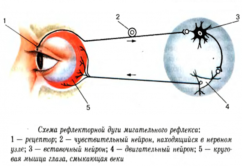
The structure of the arc of the blink reflex is also interesting. This reflex, due to its complexity, makes it possible to study the movement of excitation along an arc, which is difficult to study in other cases. The reflex arc of this reflex begins with the activation of excitatory and inhibitory neurons simultaneously. Depending on the nature of the damage, different parts of the arc are activated. The onset of the blink reflex can be triggered by the trigeminal nerve - in response to touch, auditory - in response to a sharp sound, visual - in response to a change in light or visible danger.
The reflex has early and late components. The late component is responsible for generating the response delay. As an experiment, touch the skin of the eyelid with your finger. The eye closes with lightning speed. When the skin is touched again, the reaction is slower. After the brain processes the information received, conscious inhibition of the acquired reflex occurs. Thanks to this inhibition, for example, women very quickly learn to paint their eyelids, overcoming the natural desire of the eyelid to cover the cornea of the eye.
Other variants of polysynaptic arcs are also amenable to research, but they are often too complex and not very clear to study.
No matter what heights science has reached, the blinking and knee reflexes remain the basic reflexes for studying human reactions. Studying and measuring the speed of impulse transmission in the trigeminal and facial nerves is the basis for assessing the condition of the brain stem in various pathologies and pain.
Monosynaptic reflex arc
An arc that consists of only two neurons, which is quite enough for an impulse, is called monosynaptic. A classic example of a monosynaptic arc is the knee jerk reflex. That's why detailed diagram reflex arc of the knee is located in all medical textbooks. The peculiarity of the composition of such an arc is that it does not involve the brain. The knee reflex is an unconditioned muscle reflex. In humans and other vertebrates, such muscle reflexes are responsible for survival.

It is not surprising that it is the knee reflex that is checked by a neurologist as one of the indicators of the state of the somatic nervous system. When a hammer hits a tendon, the muscle is stretched, after the irritation passes through the centripetal fiber to the spinal ganglion, the signal passes through the motor neuron to the centrifugal fiber. Skin receptors do not take part in this experiment, however, the result is very noticeable and the strength of the reaction is easy to differentiate.
The autonomic reflex arc breaks into pieces, forming a synapse, whereas in the somatic system the path covered by the impulse from the receptor to the acting skeletal muscle is not interrupted by anything.
The activity of the body is a natural reflex reaction to a stimulus. Reflex– the body’s reaction to irritation of receptors, which is carried out with the participation of the central nervous system. The structural basis of the reflex is the reflex arc.
Reflex arc- a series-connected chain of nerve cells that ensures the implementation of a reaction, a response to irritation.
The reflex arc consists of six components: receptors, afferent (sensitive) path, reflex center, efferent (motor, secretory) path, effector (working organ), feedback.
Reflex arcs can be of two types:
1) simple - monosynaptic reflex arcs (tendon reflex reflex arc), consisting of 2 neurons (receptor (afferent) and effector), with 1 synapse between them;
2) complex – polysynaptic reflex arcs. They consist of 3 neurons (there may be more) - a receptor, one or more intercalary and an effector.
The idea of a reflex arc as an expedient response of the body dictates the need to supplement the reflex arc with another link - a feedback loop. This component establishes a connection between the realized result of the reflex reaction and the nerve center that issues executive commands. With the help of this component, the open reflex arc is transformed into a closed one.
Features of a simple monosynaptic reflex arc:
1) geographically close receptor and effector;
2) reflex arc two-neuron, monosynaptic;
3) nerve fibers of group Aα (70-120 m/s);
4) short time reflex;
5) muscles contracting according to the type of single muscle contraction.
Features of a complex monosynaptic reflex arc:
1) territorially separated receptor and effector;
2) three-neuron receptor arch (there may be more neurons);
3) the presence of nerve fibers of groups C and B;
4) muscle contraction according to the tetanus type.
Features of the autonomic reflex:
1) the interneuron is located in the lateral horns;
2) the preganglionic nerve pathway begins from the lateral horns, after the ganglion - the postganglionic;
3) the efferent path of the autonomic nervous arch reflex is interrupted by the autonomic ganglion, in which the efferent neuron lies.
The difference between the sympathetic nervous arch and the parasympathetic: the sympathetic nervous arch has a short preganglionic pathway, since the autonomic ganglion lies closer to the spinal cord, and the postganglionic pathway is long.
In the parasympathetic arc, the opposite is true: the preganglionic pathway is long, since the ganglion lies close to the organ or in the organ itself, and the postganglionic pathway is short.
Topic of the lesson: Reflex, reflex arc.
Reflex(from Latin “reflexus” - reflection) - the body’s reaction to changes in the external or internal environment, carried out through the central nervous system in response to irritation of receptors.
Reflexes are manifested in the occurrence or cessation of any activity of the body: in the contraction or relaxation of muscles, in the secretion or cessation of secretion of glands, in the constriction or dilation of blood vessels, etc.
Thanks to reflex activity, the body is able to quickly respond to various changes in the external environment or its internal state and adapt to these changes. In vertebrate animals, the importance of the reflex function of the central nervous system is so great that even its partial loss (during surgical removal of certain parts of the nervous system or in case of diseases) often leads to profound disability and the inability to perform necessary vital functions without constant careful care.
The significance of the reflex activity of the central nervous system was fully revealed by the classical works of I. M. Sechenov and I. P. Pavlov. Back in 1862, I.M. Sechenov, in his epoch-defining work “Reflexes of the Brain,” stated: “All acts of conscious and unconscious life, according to their method of origin, are reflexes.”
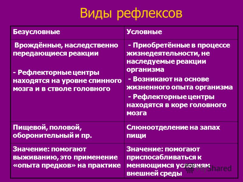
Types of reflexes
All reflex acts of the whole organism are divided into unconditioned and conditioned reflexes.
Unconditioned reflexes are inherited; they are inherent in every biological species; their arches are formed at the time of birth and normally remain throughout life. However, they can change under the influence of the disease.
Conditioned reflexes arise during individual development and accumulation of new skills. The development of new temporary connections depends on changing environmental conditions. Conditioned reflexes are formed on the basis of unconditioned ones and with the participation of higher parts of the brain.
Unconditioned and conditioned reflexes can be classified into various groups according to a number of signs.
According to biological significance
defensive
indicative
postural tonic (reflexes of body position in space)
locomotor (reflexes of body movement in space)
According to the location of the receptors, the irritation of which is caused by this reflex act
exteroceptive reflex - irritation of receptors on the external surface of the body
viscero- or interoreceptive reflex - arising from irritation of receptors of internal organs and blood vessels
proprioceptive (myotatic) reflex - irritation of receptors of skeletal muscles, joints, tendons.
According to the location of the neurons involved in the reflex
spinal reflexes - neurons located in the spinal cord
bulbar reflexes - carried out with the obligatory participation of neurons of the medulla oblongata
mesencephalic reflexes - carried out with the participation of midbrain neurons
diencephalic reflexes - neurons of the diencephalon are involved
cortical reflexes - carried out with the participation of cortical neurons cerebral hemispheres brain
Based on a number of characteristics, reflexes can be divided into groups
By type of receptor: exteroceptive (skin, visual, auditory, olfactory), interoceptive (from receptors of internal organs) and proprioceptive (from receptors of muscles, tendons, joints)
By effector: somatic or motor (skeletal muscle reflexes), for example flexor, extensor, locomotor, statokinetic, etc.; vegetative internal organs - digestive, cardiovascular, excretory, secretory, etc.
In reflex acts carried out with the participation of neurons located in the higher parts of the central nervous system, neurons located in the lower parts - in the intermediate, middle, medulla oblongata and spinal cord - always participate. On the other hand, with reflexes that are carried out by the spinal or medulla oblongata, midbrain or diencephalon, nerve impulses reach the higher parts of the central nervous system. Thus, this classification of reflex acts is to some extent arbitrary.
By the nature of the response, depending on which organs are involved in it
motor, or motor reflexes - muscles serve as the executive organ;
secretory reflexes - end with the secretion of glands;
vasomotor reflexes - manifested in the narrowing or expansion of blood vessels.
Such a classification of reflexes is conditional: if any reflex can be obtained with the preservation of one or another part of the central nervous system and the destruction of the overlying parts, this does not mean that this reflex is carried out in a normal body only with the participation of this part: in each reflex they involve to one degree or another, all parts of the central nervous system.
Any reflex in the body is carried out using a reflex arc.
Neurons and the pathways of nerve impulses during a reflex act form a so-called reflex arc:
stimulus - receptor-affector - CNS neuron - effector - response.
Reflex arc- this is the path along which irritation (signal) from the receptor passes to the executive organ. The structural basis of the reflex arc is formed by neural circuits consisting of receptor, intercalary and effector neurons. It is these neurons and their processes that form the path along which nerve impulses from the receptor are transmitted to the executive organ during the implementation of any reflex.
In the peripheral nervous system, reflex arcs (neural circuits) of the somatic nervous system are distinguished, innervating the skeletal muscles of the autonomic nervous system, innervating the internal organs: heart, stomach, intestines, kidneys, liver, etc.
According to the degree of complexity of the neural organization of reflex arcs, a distinction is made between monosynaptic, the arcs of which consist of afferent and efferent neurons (for example, knee), and polysynaptic, the arcs of which also contain 1 or more intermediate neurons and have 2 or several synaptic switches (for example, flexor).
According to the nature of the influences on the activity of the effector: excitatory - causing and enhancing (facilitating) its activity, inhibitory - weakening and suppressing it (for example, reflex increase heart rate sympathetic nerve and its reduction or cardiac arrest - vagus).
Based on the anatomical location of the central part of the reflex arcs, spinal reflexes and cerebral reflexes are distinguished. Neurons located in the spinal cord are involved in the implementation of spinal reflexes. An example of the simplest spinal reflex is the withdrawal of a hand from a sharp pin. Reflexes of the brain are carried out with the participation of neurons in the brain. Among them there are bulbar, carried out with the participation of neurons of the medulla oblongata; mesencephalic - with the participation of midbrain neurons; cortical - with the participation of neurons in the cerebral cortex.
There are monosynaptic (involving the transmission of impulses to the command neuron through one synaptic transmission) and polysynaptic (involving the transmission of impulses through chains of neurons) reflexes.
Neural organization of the simplest reflex
The simplest reflex in vertebrates is considered monosynaptic. If the arc of the spinal reflex is formed by two neurons, then the first of them is represented by a cell of the spinal ganglion, and the second is a motor cell (motoneuron) of the anterior horn of the spinal cord. The long dendrite of the spinal ganglion goes to the periphery, forming a sensitive fiber of a nerve trunk, and ends with a receptor. The axon of a neuron of the spinal ganglion is part of the dorsal root of the spinal cord, reaches the motor neuron of the anterior horn and, through a synapse, connects with the body of the neuron or one of its dendrites. The axon of the anterior horn motor neuron is part of the anterior root, then the corresponding motor nerve and ends in a motor plaque in the muscle.
Pure monosynaptic reflexes do not exist. Even the knee reflex, which is a classic example of a monosynaptic reflex, is polysynaptic, since the sensory neuron not only switches to the motor neuron of the extensor muscle, but also sends an axonal collateral that switches to the inhibitory interneuron of the antagonist muscle, the flexor muscle.
Reflex arc(nerve arc) - the path traversed by nerve impulses during the implementation of a reflex.
The reflex arc consists of:
receptor - a nerve link that perceives irritation;
afferent link - centripetal nerve fiber - processes of receptor neurons that transmit impulses from sensory nerve endings to the central nervous system;
central link - nerve center (optional element, for example for the axon reflex);
efferent link - carry out transmission from the nerve center to the effector.
effector - an executive organ whose activity changes as a result of a reflex.
There are:
monosynaptic, two-neuron reflex arcs;
polysynaptic reflex arcs (include three or more neurons).
Polysynaptic reflex arc: a nerve impulse from the receptor is transmitted along a sensory (afferent) neuron to the spinal cord. The cell body of the sensory neuron is located in the spinal ganglion outside the spinal cord. Axon of a sensory neuron in gray matter brain is connected through synapses with one or more interneurons, which, in turn, are connected to the dendrites of the motor (efferent) neuron. The axon of the latter transmits a signal from the ventral root to the effector (muscle or gland).
The reflex arc consists of five sections:
receptors that perceive irritation and respond to it with excitement. Receptors can be the endings of long processes of centripetal nerves or microscopic bodies of various shapes from epithelial cells on which the processes of neurons end. Receptors are located in the skin, in all internal organs; clusters of receptors form the sense organs (eye, ear, etc.).
sensitive (centripetal, afferent) nerve fiber transmitting excitation to the center; a neuron that has this fiber is also called sensitive. The cell bodies of sensory neurons are located outside the central nervous system - in ganglia along the spinal cord and near the brain.
the nerve center, where excitation switches from sensory neurons to motor neurons; The centers of most motor reflexes are located in the spinal cord. The brain contains centers for complex reflexes, such as protective, food, orientation, etc. In the nerve center, a synaptic connection between the sensory and motor neurons occurs.
motor (centrifugal, efferent) nerve fiber, carrying excitation from the central nervous system to the working organ; Centrifugal fiber is a long extension of a motor neuron. A motor neuron is a neuron whose process approaches the working organ and transmits a signal to it from the center.
effector - a working organ that carries out an effect, a reaction in response to stimulation of the receptor. Effectors can be muscles that contract when they receive stimulation from the center, gland cells that secrete juice under the influence of nervous stimulation, or other organs.
The simplest reflex arc can be schematically represented as formed by only two neurons: receptor and effector, between which there is one synapse. This reflex arc is called bineuronal and monosynaptic. Monosynaptic reflex arcs are very rare. An example of them is the arc of the myotatic reflex.
In most cases, reflex arcs include not two, but larger number neurons: receptor, one or more intercalary and effector. Such reflex arcs are called multineuronal and polysynaptic. An example of a polysynaptic reflex arc is the reflex of withdrawing a limb in response to painful stimulation.
Reflex arc of the somatic nervous system on the way from the central nervous system to skeletal muscle is not interrupted anywhere, unlike the reflex arc of the autonomic nervous system, which, on the way from the central nervous system to the innervated organ, is necessarily interrupted with the formation of a synapse - the autonomic ganglion.
Autonomic ganglia, depending on location, can be divided into three groups:
vertebral ganglia - belong to the sympathetic nervous system. They are located on both sides of the spine, forming two border trunks (they are also called sympathetic chains)
The prevertebral (prevertebral) ganglia are located at a greater distance from the spine, but at the same time they are located at some distance from the organs they innervate. The prevertebral ganglia include the ciliary ganglion, superior and middle cervical sympathetic nodes, solar plexus, superior and inferior mesenteric ganglia.
intraorgan ganglia are located in the internal organs: in the muscular walls of the heart, bronchi, middle and lower third of the esophagus, stomach, intestines, gallbladder, bladder, as well as in the glands of external and internal secretion. Parasympathetic fibers are interrupted on the cells of these ganglia.
This difference between the somatic and autonomic reflex arc is due to the anatomical structure of the nerve fibers that make up the neural chain and the speed of transmission of the nerve impulse through them.
For any reflex to occur, the integrity of all parts of the reflex arc is necessary. Violation of at least one of them leads to the disappearance of the reflex.
Reflex implementation scheme
In response to receptor stimulation, the nervous tissue enters a state of excitation, which is a nervous process that causes or enhances the activity of the organ. The basis of excitation is a change in the concentration of anions and cations on both sides of the membrane of the processes nerve cell, which leads to a change in the electrical potential on the cell membrane.
In a two-neuron reflex arc (the first neuron is a dorsal ganglion cell, the second neuron is a motor neuron [motoneuron] of the anterior horn of the spinal cord), the dendrite of the dorsal ganglion cell has a significant length; it follows to the periphery as part of the sensory fibers of the nerve trunks. The dendrite ends with a special device for perceiving irritation - a receptor.
Excitation from the receptor is transmitted centripetally (centripetal) along the nerve fiber to the spinal ganglion. The axon of the spinal ganglion neuron is part of the dorsal (sensitive) root; this fiber reaches the motor neuron of the anterior horn and with the help of a synapse in which signal transmission occurs using chemical substance- mediator, establishes contact with the body of the motor neuron or with one of its dendrites. The axon of this motor neuron is part of the anterior (motor) root, through which the signal travels centrifugally (centrifugally) to the executive organ, where the corresponding motor nerve ends in a motor plaque in the muscle. As a result, muscle contraction occurs.
Excitation is carried out along nerve fibers at a speed of 0.5 to 100 m/s, in isolation and does not pass from one fiber to another, which is prevented by the membranes covering the nerve fibers.
The process of inhibition is the opposite of excitation: it stops activity, weakens or prevents its occurrence. Excitation in some centers of the nervous system is accompanied by inhibition in others: nerve impulses entering the central nervous system can delay certain reflexes.
Both processes - excitation and inhibition - are interconnected, which ensures coordinated activity of organs and the entire organism as a whole. For example, during walking, contraction of the flexor and extensor muscles alternates: when the flexion center is excited, impulses follow to the flexor muscles, at the same time, the extension center is inhibited and does not send impulses to the extensor muscles, as a result of which the latter relax, and vice versa.
The relationship that determines the processes of excitation and inhibition, i.e. self-regulation of body functions is carried out using direct and feedback connections between the central nervous system and the executive organ. Feedback (“reverse afferentation” according to P.K. Anokhin), i.e. connection between executive body and the central nervous system, implies the transmission of signals from the working organ to the central nervous system about the results of its work at any given moment.
Complete the task:
A. When pressing on eyeballs within 10–15 s the human heart rate slows down. Establish the sequence of those parts of the reflex arc that participate in this reflex by selecting the necessary elements:
1.heart; 2. afferent neuron; 3.vagus nerve; 4.cerebellum; 5. medulla oblongata; 6.sympathetic nerve; 7.mechanoreceptors of the eye.
B. Establish the sequence of passage of a nerve impulse along the reflex arc of a protective reaction to enhance heat transfer in a person, selecting the necessary elements from those proposed:
1.mechanoreceptors of the ciliary epithelium of the skin; 2. intercalary neurons of the medulla oblongata; 3. afferent neuron; 4.efferent neuron; 5.smooth muscles of the skin relax; 6. thermoreceptors of the dermis; 7. interneurons of the hypothalamus; 8. The lumen of the capillaries expands.
B. Establish the sequence of passage of a nerve impulse along the reflex arc of the protective sneezing reflex in a person, selecting the necessary elements from those proposed:
1. mechanoreceptors of the ciliated epithelium of the nasal cavity; 2. interneurons of the medulla oblongata; 3.efferent neuron; 4. afferent neuron; 5.effector; 6. interneurons of the midbrain; 7. mechanoreceptors of the larynx.
D. If you touch the lips of a sleeping child with a pacifier, then he makes sucking movements. Establish the sequence of passage of a nerve impulse along the reflex arc by selecting the necessary elements:
1. efferent neuron; 2.chemoreceptors of the lips; 3. diencephalon; 4. medulla oblongata; 5. afferent neuron; 6. orbicularis oris muscle, tongue; 7. mechanoreceptors of the lips; 8.cerebral cortex.
D. If the stomach is poisoned or the stomach is full of food, vomiting may occur. Indicate the sequence... taking part in this reflex, selecting the necessary: 1. thermoreceptors of the lips; 2.stomach receptors; 3. diencephalon; 4.efferent neuron; 5. medulla oblongata; 6. afferent neuron; 7.muscles of the stomach.
E. . Establish the sequence of passage of a nerve impulse when a conditioned salivary reflex occurs in a person, selecting the necessary elements from those proposed:
1.type of lemon; 2. medulla oblongata; 3.receptors of the retina; 4.cerebral cortex; 5. afferent neuron; 6. efferent neuron; 7. increased secretion of the salivary glands; 8. chemoreceptors of the tongue.
E. Establish the sequence of transmission of a nerve impulse along the reflex arc of the human parasympathetic nervous system, selecting the necessary elements from those proposed:
1.preganglionic neuron; 2. postganglionic neuron; 3. smooth muscles of the bladder; 4.mechanoreceptors of the bladder; 5.sympathetic trunk; 6.spinal cord; 7.sensitive neuron; 8.cerebral cortex.