The human brain consists of white and gray matter. The first is everything that is filled between the gray matter on the cortex and on the surface there is a uniform layer of gray matter with nerve cells, the thickness of which is up to four and a half millimeters.
Let's study in more detail what gray and white matter is in the brain.
What are these substances made of?
The substance of the central nervous system is of two types: white and gray.
White matter consists of many nerve fibers and processes nerve cells, the shell of which is white.
Gray matter consists of processes. Nerve fibers connect different parts of the central nervous system and nerve centers.
Gray and white matter of the spinal cord
The heterogeneous substance of this organ is gray and white. The first is formed by a huge number of neurons, which are concentrated in nuclei and come in three types:
- radicular cells;
- tufted neurons;
- internal cells.
The white matter of the spinal cord surrounds gray matter. It includes nerve processes that make up three fiber systems:
- intercalary and afferent neurons connecting different parts of the spinal cord;
- sensory afferents, which are long centripetal;
- motor afferent or long centrifugal.

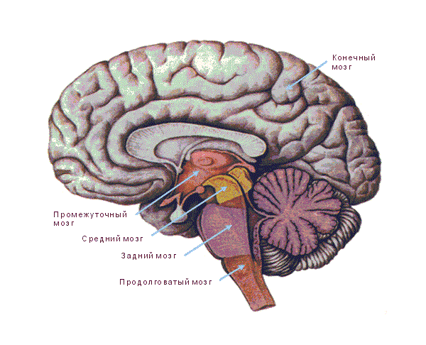
Medulla oblongata
From the anatomy course we know that the spinal cord passes into the medulla oblongata. The part of this brain at the top is thicker than at the bottom. Its average length is 25 millimeters, and its shape resembles a truncated cone.
It develops gravitational and auditory organs associated with breathing and blood circulation. Therefore, the nuclei of gray matter here regulate balance, metabolism, blood circulation, breathing, and coordination of movements.
hindbrain
This brain consists of the pons and the cerebellum. Let's look at the gray and white matter in them. The bridge is a large white ridge on the back side of the base. On the one hand, its border with the cerebral peduncles is pronounced, and on the other, with the medulla oblongata. If you make a cross section, the white matter of the brain and the gray nucleus will be visible very clearly. Transverse fibers divide the bridge into ventral and dorsal sections. In the ventral part, the white matter of the pathways is mainly present, and the gray matter forms its nuclei here.
The dorsal part is represented by nuclei: switching, sensory systems and cranial nerves.
The cerebellum is located under the occipital lobes. It includes the hemispheres and the middle part called the “worm”. Gray matter makes up the cerebellar cortex and nuclei, which are tent-shaped, spherical, corky and dentate. The white matter of the brain in this part is located under the cerebellar cortex. It penetrates into all gyri as white plates and consists of different fibers that either connect the lobules and gyri, or are directed to the internal nuclei, or connect sections of the brain.

Midbrain
It starts from the mesencephalon. On the one hand, it corresponds to the surface of the brain stem between and the superior medullary velum, and on the other, to the area between the mammillary bodies and the anterior part of the pons.
It includes the cerebral aqueduct, on one side of which the boundary is provided by the roof, and on the other by the covering of the cerebral peduncles. In the ventral area, the posterior perforated substance and legs are distinguished big brain, and on the dorsal side - the plate of the roof and the handles of the lower and upper tubercles.
If we look at the white and gray matter of the brain in the cerebral aqueduct, we will see that the white surrounds the central gray matter, consisting of small cells and having a thickness of 2 to 5 millimeters. It consists of the trochlear, trigeminal and oculomotor nerves, together with the accessory nucleus of the latter and the intermediate nucleus.
Diencephalon
It is located between the corpus callosum and the fornix, and on the sides it fuses with the Dorsal section consists of the visual tuberosities, on the upper part of which there is the epitubercle, and in the ventral part there is the inferior tuberosity region.
The gray matter here consists of nuclei that are associated with centers of sensitivity.
White matter is represented by pathways different directions, guaranteeing the connection of formations with the cerebral cortex and nuclei. The diencephalon also includes the pituitary gland and pineal gland.

Finite brain
It is represented by two hemispheres, which are separated by a gap running along them. It is connected in depth by the corpus callosum and commissures.
The cavity is represented by the lateral ventricles located in one and the second hemisphere. These hemispheres consist of:
- a cloak of neocortex or six-layer cortex, distinguished by nerve cells;
- striatum from the basal ganglia - ancient, old and new;
- partitions.
But sometimes there is another classification:
- olfactory brain;
- subcortex;
- gray matter of the cortex.
Without touching on the gray matter, let's focus immediately on the white matter.

On the characteristics of the white matter of the hemispheres
The white matter of the brain occupies all the space between the gray and basal ganglia. There is a huge number of nerve fibers here. The white matter contains the following areas:
- central substance of the internal capsule, corpus callosum and long fibers;
- radiant crown of radiating fibers;
- semi-oval center in outer parts;
- a substance found in the convolutions between the furrows.
Nerve fibers are:
- commissural;
- associative;
- projection.
The white matter includes nerve fibers that are connected by the convolutions of one and the other cerebral cortex and other formations.
Nerve fibers

Commissural fibers are mainly found in the corpus callosum. They are located in the cerebral commissures, which connect the cortex on different hemispheres and symmetrical points.
Association fibers group areas on one hemisphere. In this case, the short ones connect neighboring convolutions, and the long ones connect those located at a far distance from each other.
Projection fibers connect the cortex with those formations located below, and then with the periphery.
If the internal capsule is viewed in section from the front, the lenticular nucleus and the posterior limb will be visible. Projection fibers are divided into:
- fibers located from the thalamus to the cortex and in the opposite direction, they excite the cortex and are centrifugal;
- fibers directed to the motor nuclei of the nerves;
- fibers that conduct impulses to the muscles of the whole body;
- fibers directed from the cortex to the pontine nuclei, providing a regulatory and inhibitory effect on the work of the cerebellum.
Those projection fibers that are located closest to the cortex create the corona radiata. Then their main part passes into the internal capsule, where the white matter is located between the caudate and lenticular nuclei, as well as the thalamus.
On the surface there is extremely complex drawing, where grooves and ridges alternate between them. They are called convolutions. Deep grooves divide the hemispheres into large areas called lobes. In general, the grooves of the brain are deeply individual; they can differ greatly from person to person. different people.
The hemispheres have five lobes:
- frontal;
- parietal;
- temporal;
- occipital;
- island.
The central sulcus originates at the top of the hemisphere and moves down and forward to the frontal lobe. The area posterior to the central sulcus is the parietal lobe, which ends in the parieto-occipital sulcus.
Frontal lobe is divided into four convolutions, vertical and horizontal.
The lateral surface is represented by three convolutions, which are delimited from each other.
The furrows of the occipital lobe are variable. But everyone, as a rule, has a transverse one, which is connected to the end of the interparietal groove.
On the parietal lobe there is a groove that runs horizontally parallel to the central one and merges with another groove. Depending on their location, this lobe is divided into three convolutions.
The island has a triangular shape. It is covered with short convolutions.
Brain lesions

Thanks to the achievements modern science High-tech brain diagnostics have become possible. Thus, if there is a pathological focus in the white matter, it can be detected at an early stage and therapy can be prescribed in a timely manner.
Among the diseases that are caused by damage to this substance are its disorders in the hemispheres, pathologies of the capsule, corpus callosum and syndromes of a mixed nature. For example, if the hind leg is damaged, one half of the human body can be paralyzed. This problem may develop with sensory disturbances or visual field defects. Malfunctions of the corpus callosum lead to mental disorders. In this case, the person ceases to recognize surrounding objects, phenomena, etc., or does not perform purposeful actions. If the lesion is bilateral, swallowing and speech disorders may occur.
The importance of both gray and white matter in the brain cannot be overstated. Therefore, the earlier the presence of pathology is detected, the greater the chance that treatment will be successful.
The brain consists of gray and white matter. White matter occupies the entire space between the gray matter of the cerebral cortex and the basal ganglia. The surface of the hemisphere, the cloak (pallium), is formed by a uniform layer of gray matter 1.3 - 4.5 mm thick, containing nerve cells.
First, let's look at white matter.
White matter has four parts:
1) the central substance of the corpus callosum, internal capsule and long associative fibers.
2) radiant crown (corona radiata), formed by radiating fibers entering and leaving the internal capsule (capsula interna);
3) the area of white matter in the outer parts of the hemisphere - the semi-oval center (centrum semiovale);
4) white matter in the gyri between the sulci;
Nerve fibers of white matter are divided into projection, associative and commissural.
The white matter of the hemispheres is formed by nerve fibers connecting the cortex of one gyrus with the cortex of other gyri of its and the opposite hemispheres, as well as with underlying formations.
Two brain commissures, commissura anterior and commissura fornicis, are much smaller in size and relate to the olfactory brain of the rhinencephalon and connect: commissura anterior - olfactory lobes and both parahippocampal gyri, commissura fornicis - hippocampi.
Most of the commissural fibers are part of the corpus callosum, which connects the parts of both hemispheres belonging to the brain.
Commissural fibers, which are part of the cerebral commissures, or commissures, connect not only symmetrical points, but also the cortex belonging to different lobes of the opposite hemispheres.
Association fibers connect different parts of the cortex of the same hemisphere.
Associative fibers are divided into short and long.
Short fibers connect neighboring convolutions in the form of arcuate bundles.
Long association fibers connect areas of the cortex that are more distant from each other.
Projection fibers connect the cerebral cortex with the underlying formations, and through them with the periphery. These fibers are divided into centripetal (ascending, corticopetal, afferent).
On a frontal section of the brain, the internal capsule looks like an oblique white stripe that continues into the cerebral peduncle.
In the internal capsule, the anterior leg (crus anterius) is distinguished - between the caudate nucleus and the anterior half of the inner surface of the lentiform nucleus, hind leg(crus posterius), - between the thalamus and the posterior half of the lenticular nucleus and genu (genu), lying at the inflection point between both parts of the internal capsule. Projection fibers can be divided according to their length into the following three systems, starting with the longest:
1. Fibrae thalamocorticalis et corticothalamici - fibers from the thalamus to the cortex and back from the cortex to the thalamus. Conducting excitation towards the cortex, and centrifugal (descending, corticofugal, efferent).
2. Tractus corticonuclearis - pathways to the motor nuclei of the cranial nerves. Since all motor fibers are collected in a small space in the internal capsule (the knee and the anterior two-thirds of its posterior leg), if they are damaged in this place, unilateral paralysis of the opposite side of the body is observed.
3. Tractus corticospinalis (pyramidalis) conducts motor volitional impulses to the muscles of the trunk and limbs.
4. Tractus corticopontini - paths from the cerebral cortex to the pontine nuclei. Using these pathways, the cerebral cortex has an inhibitory and regulatory effect on the activity of the cerebellum.
Projection fibers in the white matter of the hemisphere closer to the cortex form the corona radiata, and then the main part of them converges into the internal capsule, which is a layer of white matter between the lentiform nucleus (nucleus lentiformis) on one side, and the caudate nucleus (nucleus caudatus) and thalamus ( thalamus) - on the other.
Now let's look at the gray matter.
The surface of the cloak has a very complex pattern, consisting of alternating various directions grooves and ridges between them, called convolutions, gyri.
Deep permanent grooves are used to divide each hemisphere into large areas called lobes, lobi; the latter, in turn, are divided into lobules and convolutions.
The size and shape of the grooves are subject to significant individual fluctuations, as a result of which not only the brains of different people, but even the hemispheres of the same individual are not quite similar in the pattern of the grooves.
There are five lobes of the hemisphere: frontal (lobus frontalis), parietal (lobus parietalis), temporal (lobus temporalis), occipital (lobus occipitalis) and a lobe hidden at the bottom of the lateral sulcus, the so-called insula.
The central sulcus (sulcus cenrtalis) begins at the upper edge of the hemisphere and goes forward and down. The area of the hemisphere located in front of the central sulcus. Refers to the frontal lobe. The portion of the brain surface posterior to the central sulcus constitutes the parietal lobe. The posterior border of the parietal lobe is the end of the parieto-occipital sulcus (sulcus parietooccipitalis), located on the medial surface of the hemisphere.
Frontal lobe. In the posterior region outer surface This lobe runs sulcus precentralis almost parallel to the direction of sulcus centralis. Two grooves run from it in the longitudinal direction: sulcus frontalis superior et sulcus frontalis inferior. Due to this, the frontal lobe is divided into four convolutions.
The vertical gyrus, gyrus precentralis, is located between the central and precentral sulci. The superior lateral surface of the hemisphere is delimited into lobes by three sulci: the lateral, central and the upper end of the parieto-occipital sulcus.
The lateral groove (sulcus cerebri lateralis) begins on the basal surface of the hemisphere from the lateral fossa and then passes to the superolateral surface
The lobe consists of a number of convolutions, called in some places lobules, which are limited by the grooves of the brain surface.
The horizontal convolutions of the frontal lobe are: superior frontal (gyrus frontalis superior), middle frontal (gyrus frontalis medius) and inferior frontal (gyrus frontalis inferior).
Temporal lobe. The lateral surface of this lobe has three longitudinal convolutions, delimited from each other by sulcus temporalis superior and sulcus temporalis inferior. The gyrus temporalis medius extends between the superior and inferior temporal grooves. Below it runs the gyrus temporalis inferior.
Occipital lobe. The grooves on the lateral surface of this lobe are variable and inconsistent. Of these, the transversely running sulcus occipitalis transversus is distinguished, usually connecting to the end of the interparietal sulcus.
Parietal lobe. On it, approximately parallel to the central groove, there is a sulcus postcentralis, usually merging with the sulcus intraparietalis, which runs in a horizontal direction. Depending on the location of these grooves, the parietal lobe is divided into three gyri.
The vertical gyrus, gyrus postcentralis, runs behind the central sulcus in the same direction as the precentral gyrus. Above the interparietal sulcus is the superior parietal gyrus, or lobule (lobulus parietalis superior), below - lobulus parietalis inferior.
Island. This lobe has the shape of a triangle. The surface of the insula is covered with short convolutions.
The lower surface of the hemisphere in that part that lies anterior to the lateral fossa belongs to the frontal lobe.
On the posterior portion of the basal surface of the hemisphere, two grooves are visible: the sulcus occipitotemporalis, running in the direction from the occipital pole to the temporal and limiting the gyrus occipitotemporalis lateralis, and the sulcus collateralis running parallel to it. Here the sulcus olfactorius runs parallel to the medial edge of the hemisphere. Parallel to and above this groove, the sulcus cinguli runs along the medial surface of the hemisphere. Between them is the gyrus occipitotemporalis medialis.
There are two gyri located medially from the collateral sulcus: between the posterior section of this sulcus and the sulcus calcarinus lies the gyrus lingualis; between the anterior section of this groove and the deep sulcus hippocampi lies the gyrus parahippocampalis.
The gyrus adjacent to the brain stem is already located on the medial surface of the hemisphere.
Behind the precuneus lies a separate area of the cortex belonging to the occipital lobe - the cuneus. Between the lingual sulcus and the sulcus of the corpus callosum stretches the cingulate gyrus (gyrus cinguli), which, through the isthmus (isthmus), continues into the parahippocampal gyrus, ending with the hook (uncus). Gyrus cinguli, isthmus and gyrus parahippocampalis form together a vaulted gyrus (gyrus fornicatus), which describes an almost complete circle, open only below and in front.
On the medial surface of the hemisphere there is a groove of the corpus callosum (sulcus corpori callosi), running directly above the corpus callosum and continuing with its posterior end into the deep sulcus hippocampi, which is directed forward and downward.
The paracentral lobule (lobulus paracentralis) is a small area above the lingual sulcus. From the paracentral lobule there is quadrangular surface(the so-called precuneus, precuneus). It belongs to the parietal lobe. The vaulted gyrus is not related to any of the cloak lobes. It belongs to the limbic region. The limbic region is part of the neocortex of the cerebral hemispheres, occupying the cingulate and parahippocampal gyri; part of the limbic system.
Pulling apart the edge of the sulcus hippocampi, one can see a narrow jagged gray stripe, which is a rudimentary gyrus of the gyrus dentatus.
References
1. M.G. Prives, N.K. Lysenkov, V.I. Bushkovich. Human anatomy. M., 1985
2. Great medical encyclopedia. vol. 11, M., 1979
3. Great medical encyclopedia. vol. 6, M., 1977
|
Gray and white matter of the brain Abstract. How to treat the disease? Gray and white matter of the brain Abstract. Traditional methods of treatment and healing. Unique healing video sessions. |
Brain tissue is made up of nerve cells (neurons). Their collection is called the gray and white matter of the brain. In the first case, there is a concentration of neuron bodies, and in the second, their axons (processes). The gray matter of the brain is its outer layer. Its volume actually reaches half a centimeter. White is located inside this meninges. However, in the spinal cord the opposite is true.
To fully understand the characteristics of the matter that makes up the brain and spinal cord, it is necessary to study its anatomical details. You can see the white and gray matter in this image:
You can see the gray and white matter of the spinal cord in this picture: 
The substance that makes up brain tissue has following features buildings:
- The light part. From Latin it is translated as substantia alba and represents important component CNS (central nervous system). White matter consists primarily of neuronal processes covered with myelin, called axons. Substantia alba gets its color from the myelin layer. In the brain tissues of the head, the substance is located inside the gray matter (substantia grisea). The structure of the spinal cord is somewhat different from the brain. In it, the white matter is outside the gray, and it should form the lateral, posterior and anterior cords. The only place where the substantia alba in the head is around the substantia grisea area is in the nuclei (ganglia);
- The dark part. The gray matter of the brain is formed from the cell bodies of neurons, capillaries, and neuropil. The substance gets its color from small blood vessels. It is located in the departments responsible for muscle tissue, perception, memory, emotions and speech.
Spinal cord
The spinal cord is fundamentally different in structure from the brain. In it, light and dark substance is concentrated in nuclei, which are of the following types:
- Internal;
- Beam;
- Radicular.
Unlike the brain tissues of the head, in the back the substantia alba is located outside the substantia grisea. Among other features, the components of the white matter of the spinal cord can be distinguished:
- Intercalary and afferent neurons that serve to connect various parts spinal cord;
- Afferent neurons (sensitive);
- Motor neurons.

Medulla oblongata
The spinal cord passes directly into the medulla oblongata (myelencephalon). Its size usually does not exceed 2-3 cm, and in its own way appearance this section looks like a truncated cone. He is primarily responsible for the following functions:
- Circulation;
- Respiratory system;
- Equilibrium;
- Coordination of movements;
- Exchange processes.
Posterior brain tissue
Directly above the medulla oblongata is the pons, and to the right is the cerebellum. The first section is presented in the form of a light-colored roller. It is associated with the cerebral peduncles and myelencephalon.
Transverse fibers divide the bridge into the following parts:
- Ventral (gastric). In this area, the substantia alba is represented predominantly by conductive fibers, and the substantia grisea has its nuclei here;
- Dorsal (dorsal). It consists of the following elements:
- Switch cores;
- Network formation;
- Sensory systems;
- Nerve pathways.
The cerebellum is located just below the occipital part of the brain. It consists of 2 hemispheres and a middle part. Gray matter is presented in the form of nuclei (dentate, cork-shaped, spherical, tent-shaped) and cortex. The white substance is under the dark shell. It is located in all convolutions and mainly consists of fibers that perform the following purposes:
- Connect the cerebral lobes and gyri;
- They follow the nuclei localized inside;
- Link departments.
Central brain tissue
The middle section is localized between the epiphysis and the cover like a sail. Next to it is the mastoid body and the bridge. On the gastric portion of the central brain tissue, a perforated substance can be seen, and on the dorsal part, the upper and lower sides of the tubercles.
The gray and white matter of the brain in this section has its own characteristics. The light substance predominantly surrounds the dark substance, which consists of paired cranial nerves.
Intermediate tissues
The intermediate section is located next to the fornix and corpus callosum. With its sides it connects with the anterior medulla (terminus). The dorsal part of the intermediate tissues consists of tubercles responsible for vision. The supratuberculum lies above them, and the inferior tubercular part is localized in the gastric system. The diencephalon also includes the pituitary gland and pineal gland.
Substantia grisea is presented in this place in the form of nuclei, which are directly connected to the sensitive centers. Substantia alba is a conductive pathway. The purpose of the latter is to connect formations with the surface of the brain and its nuclei.

Forebrain tissues
The anterior section is also called the terminal section. It consists of two hemispheres separated by a depression. It runs along the entire section and connects below with the corpus callosum. In the cavity of the telencephalon tissues there are lateral ventricles, and the hemispheres themselves consist of the following components:
- Isocortex;
- Striatum;
- Partitions.
The gray matter in the anterior region forms the cerebral cortex and basal ganglia. The white substance takes up all the space between them.
It plays the role of conducting pathways, which are divided into 3 groups:
- Associative. This type of fiber serves to connect different parts of the cortex in the region of the 1st hemisphere. There are short and long associative paths. The first type is presented as an arc-shaped accumulation of substance. It connects parts of the cortex of adjacent gyri. Long paths connect the lobes of the hemispheres;
- Commissural. They are localized in brain adhesions and are responsible for connecting formations in both hemispheres. The basis of the commissural fibers is the corpus callosum. Parts of this formation connect the gray matter of certain lobes to each other;
- Projection. The fibers of this group form the capsule and corona radiata. The first formation is a plate of white matter. It is surrounded by the lenticular and caudate nuclei and the hypothalamus. The capsule itself contains 2 legs and a knee. Fibers localized closer to the cortex form the corona radiata. The role of these pathways is to connect the cortex with the formations below.
Surface of the brain
On the surface of the brain (cortex) you can see a rather interesting and complex pattern. From an anatomical point of view, the alternation of grooves and ridges is clearly visible. The latter are located between them and are called convolutions.
The grooves are depressions and divide the hemispheres into certain parts called lobes. You can see them in this picture:
 The size of the grooves and medullary lobes is most often individual and differences may be observed in each person. However there is certain standards, which experts focus on:
- Central groove. It begins on the superior surface of the hemispheres and separates the parietal and frontal lobes. On the sides of it remain the temporal parts;
- Frontal lobe. It includes 4 convolutions and this area borders the parietal and temporal parts;
- Temporal. It consists of 3 convolutions separated from each other. Border this area with all other shares;
- Occipital lobe. In many people it differs in the structure of the grooves, but in most cases the transverse depression is associated with the interparietal one. This lobe borders the temporal and parietal;
- Parietal. It includes three convolutions and borders this area with all the others.
The surface of the brain is represented by gray matter and you can see this in this figure:

Damage to white or gray matter
IN recent years Medicine has advanced significantly and current technologies make it possible to scan brain tissue for the presence of pathological processes. If damage is detected in the white or gray matter, then a course of therapy can be started immediately. In this case, the chances of completely eliminating the problem will be much greater.
Depending on the location, damage to the substance is possible various options symptoms. If the posterior cerebral peduncle is injured, the patient may experience partial paralysis. Against the background of this phenomenon, vision problems and deterioration in sensitivity often occur. If the corpus callosum is damaged, mental disturbances are possible. Gradually, a person may stop recognizing people close to him and even ordinary objects. In the presence of a bilateral focus, problems with swallowing and speech defects are added to the symptoms.
Brain tissue is a collection of white and gray matter. Each of them is responsible for certain vital functions. If one of the substances is damaged, a person can die or become disabled, so it is important to promptly detect the presence of pathological processes using modern diagnostic methods.
30.10.2013
The white matter of the brain consists of large number nerve fibers that fill the space between the cerebral cortex and the basal ganglia. They spread in different directions and form pathways cerebral hemispheres. Conventionally, nerve fibers are divided into three groups: associative, commissural (transverse), projection.
Associative
They realize the relationship between different zones of the cortex localized in one hemisphere. There are short ones that connect neighboring convolutions with each other, and long ones that connect distant areas. The short ones, lying directly under the cortex, are called subcortical, and those located in the deep layers are called intracortical. Long ones include, for example, the upper and lower longitudinal beams. The superior longitudinal fasciculus originates in the frontal lobe and penetrates through the occipital lobe into the temporal lobe. The inferior unites the temporal and occipital lobes. In addition, the uncinate fasciculus is located between the temporal and frontal lobes. Another formation is the belt, which consists of fibers in the lumbar gyrus, the function of which is to connect the subcallosal body and the hook.
Commissural
They are part of the brain commissures (commissures), connecting the symmetrical areas of the hemispheres. Therefore, they have a common transverse orientation. Thanks to these fibers, the possibility of combining their functions is realized. They form three cerebral commissures, the most massive of which is the corpus callosum. It consists of the largest number transverse fibers that connect the neocortex with the corresponding zones of the opposite hemisphere. The anterior commissure links together the two olfactory bulbs and the frontal lobe. The fornix is formed by arcuate fascicles located between the hippocampus and mastoid bodies.
Projection
Connect cerebral cortex with underlying parts of the central nervous system. They are combined into a semi-oval center (corona radiata), which is immersed in the white matter of the brain. There are afferent (carrying, centripetal) pathways, which transmit impulses from organs and tissues of the body to the brain, and efferent (carrying, centrifugal) projection pathways, which transmit excitation from the central nervous system.
Between the optic thalamus and the basal ganglia there is a cluster of projection fibers in the form of a curved plate of white matter, which is called the internal capsule. It consists of the following sections: anterior leg, knee, posterior leg. Each of the elements of the internal capsule is formed by paths and bundles. For example, the anterior leg is formed by the anterior thalamic radiates, which mediate the connection between the nuclei of the thalamus and the frontal lobe, and the frontal-pontine tract, which connects the frontal lobe and the pontine nuclei. The knee of the internal capsule serves as the point of contact for both legs. It forms the corticonuclear tract, which in turn is an integral part of the pyramidal tract and tends to the nuclei of the cranial nerves. The posterior leg is represented by the following fibers: corticospinal, corticorangular, corticoreticular, corticothalamic, thalamo-parietal, central thalamic radiates, connecting the corresponding elements of the brain.
Functions of white matter of the brain
The white matter of the cerebral hemispheres provides interconnection between different parts of the nervous system. This allows her to coordinate all the work of our body.
The white matter of the brain connects homologous elements of both hemispheres.
Realizes the connection between the visual thalamus and the cortical areas.
Connects areas of the cortexcerebral hemispheres with other parts of the nervous system.
Forms close relationships between the gyri within the right as well as the left hemispheres.
Defeatwhite matter of the brain
Among the diseases affecting the white matter of the brain, limited pathologies of the internal capsule, disorders of the substance of the hemispheres, pathologies of the corpus callosum, and mixed syndromes are distinguished.
If the knee and the anterior portion of the posterior leg are damaged, hemiplegia develops -paralysis of the muscular system of one half of the human body.
Damage to the posterior part of this leg is accompanied by sensory disturbances and the “three hemi syndrome”: hemianesthesia (loss of pain and temperature sensitivity of half the face on one side, the trunk and limbs on the opposite), hemianopsia (visual field defect) and hemiataxia (impaired proprioceptive sensitivity).
Defects white matter of the hemispheres are accompanied by symptoms close to those described above; in addition, a complete half nature of the pathology may occur.
Damage to the corpus callosum provokes disorders of the patient’s mental functions. For example, agnosia (failure to recognize phenomena and objects), apraxia (lack of purposeful actions) may occur, and pseudobulbar signs are also typical.
Bilateral lesions present with speech and swallowing disorders and pyramidal symptoms.
In the brain, gray and white matter are distinguished, but their distribution here is much more complex than in the spinal cord. Most of the gray matter of the brain is located on the surface of the cerebrum and cerebellum, forming their cortex. The smaller part forms numerous subcortical nuclei, surrounded by white matter. All gray matter nuclei consist of multipolar neurons.
Gray matter contains the cell bodies of neurons, from which the nuclei of the central nervous system and cortex are formed. White matter consists of processes of neurons that form bundles and tracts that are components of the pathways of the central nervous system.
White matter in the brain occupies all the space between the gray matter of the cerebral cortex and the basal ganglia. The surface of the hemisphere, the mantle, is formed by a uniform layer of gray matter 1.3–4.5 mm thick, containing nerve cells.
White matter has four parts:
central substance of the corpus callosum, internal capsule and long associative fibers;
radiant crown (corona radiata), formed by radiating fibers entering and leaving the internal capsule;
the area of white matter in the outer parts of the hemisphere - the semi-oval center;
white matter in the gyri between the sulci.
Nerve fibers of white matter are divided into projection, associative and commissural.
The white matter of the hemispheres is formed by nerve fibers connecting the cortex of one gyrus with the cortex of other gyri of its and the opposite hemispheres, as well as with underlying formations.
Two brain commissures, commissura anterior and commissura fornicis, are much smaller in size, belong to the olfactory brain and connect: commissura anterior - olfactory lobes and both parahippocampal gyri, commissura fornicis - hippocampi.
Commissural fibers, which are part of the cerebral commissures, or commissures, connect not only symmetrical points, but also the cortex belonging to different lobes of the opposite hemispheres. Association fibers connect different parts of the cortex of the same hemisphere. They are divided into short and long fibers.
Short fibers connect neighboring convolutions in the form of arcuate bundles. Long associative fibers connect areas that are more distant from each other.
The internal capsule is a thick, angled plate of white matter, bounded on the lateral side by the lenticular nucleus, and on the medial side by the head of the caudate nucleus and the thalamus. The internal capsule is formed by projection fibers connecting the cerebral cortex with other parts of the central nervous system. Fibers of ascending pathways. Diverging in different directions towards the cortex of the hemisphere, they form a corona radiata. Downward, the fibers of the descending pathways of the internal capsule in the form of compact bundles are directed to the peduncle of the midbrain. On a frontal section of the brain, the internal capsule looks like an oblique white stripe that continues into the cerebral peduncle. In the internal capsule, the anterior leg is distinguished - between the caudate nucleus and the anterior half of the inner surface of the lentiform nucleus, as well as the posterior leg - between the thalamus and the posterior half of the lenticular nucleus and the knee. Projection fibers according to their length can be divided into the following three systems:
Fibrae thalamocorticalis et corticothalamici - fibers from the thalamus to the cortex and back from the cortex to the thalamus; conducting excitation towards the cortex and centrifugal (efferent).
Tractus corticonuclearis - pathways to the motor nuclei of the cranial nerves.
Tractus corticospinalis (pyramidalis) - conducts motor volitional impulses to the muscles of the trunk and limbs.
Tractus corticopontini - pathways from the cerebral cortex to the pontine nuclei. Using these pathways, the cerebral cortex has an inhibitory and regulatory effect on the activity of the cerebellum.
Projection fibers in the white matter of the hemisphere closer to the cortex form the corona radiata, and then the main part of them converges into the internal capsule.
Medulla oblongata.
It contains the nuclei of gray matter related to balance, coordination of movements, as well as the regulation of metabolism, respiration and blood circulation.
The gray matter of the medulla oblongata is represented by the following nuclei:
1) The olive nucleus (nucleus olivaris) has the appearance of a convoluted plate of gray matter, protruding out of the medulla oblongata. Lies in an olive tree. Responsible for balance.
2) Reticular formation (formatio reticularis), formed from the interweaving of nerve fibers and nerve cells lying between them. It contains the respiratory and vascular centers. Responsible for maintaining posture and locomotion (static vestibular reflexes), and performing protective functions (coughing, sneezing, vomiting)
3) Nuclei of four pairs of lower cephalic nerves (XII – IX).
4) The nuclei of the vagus nerve are the centers of respiration and blood circulation.
5) Wedge-shaped and thin nuclei - switching nuclei.
The white matter of the medulla oblongata contains long and short fibers. The long ones include the descending pyramidal tracts passing into the anterior cords of the spinal cord. In the nuclei of the posterior funiculi there are bodies of the second neurons of the ascending sensory pathways. In the medulla oblongata there are two intersections of long pathways: the ventral motor and dorsal sensory.
TO shortcuts These include bundles of nerve fibers that connect individual nuclei of the gray matter, as well as the nuclei of the medulla oblongata with neighboring parts of the brain.
Hindbrain.
Consists of two parts: the pons and the cerebellum. The pons contains longitudinal and transverse fibers, between which the own nuclei of the gray matter are scattered. Longitudinal fibers belong to the pyramidal tracts, which are connected to the pons proper nuclei, from where the transverse fibers that go to the cerebellar cortex originate. The surface of the cerebellum is covered by a layer of gray matter that makes up the cerebellar cortex. The cortex has three layers:
1 external, or molecular - contains various cellular elements, but few neurons. Consists mainly of interwoven basilar fibers, i.e. unmyelinated, and contains a small number of irregularly scattered small cell nuclei. It contains parallel fibers and many dendrites of Purkinje cells. Basket neurons and stellate neurons are also located here.
2 ganglion - contains large pear-shaped cells (Purkinje cells), located in 1 row. One axon extends from each such cell, going deep into the cerebellum, and the dendrites form a tree above the cell, their branching is perpendicular to the convolutions. These dendrites are equipped with spines.
3 granular, or granular - contains many granular cells. These are the smallest neurons. Their body is mainly occupied by the nucleus, around which there is a narrow layer of protoplasm. There are also two types of Golgi cells: short-axon and long-axon. The former are involved in the formation of the cerebellar lumbules, and the latter, entering the white matter of the cerebellum, connect various areas of its cortex. Behind the granular layer is the white matter, which contains the subcortical nuclei. Highlight:
Globular nucleus (nucleus globosus)
Corky nucleus (n. emboliformis)
Tent core (n.fastigii)
Dentate nucleus (n.dentatus).
The cerebellum has two types of afferent fibers: mossy and liana-shaped. The cerebellar fibers form three pairs of peduncles:
Lower legs (to the medulla oblongata)
Middle legs (toward bridge)
Upper legs (to the roof of the midbrain).
Midbrain.
The midbrain consists of a quadrigeminal colliculus, which includes 2 pairs of superior and inferior colliculi. The midbrain also includes 2 pairs of hillocks. The superior colliculi contain the visual nuclei, and the inferior colliculi contain the auditory nuclei. It contains red nuclei and substantia nigra, which belong to a system of fibers that does not pass through the pyramid of the medulla oblongata. They regulate automatic unconscious movements. The substantia nigra secretes the hormone dopamine, which suppresses excessive activity of the motor nuclei of the telencephalon. The central gray matter contains the nuclei of the III and IX cranial nerves.
The white matter of the midbrain makes up the descending tract, connecting the red nuclei and the anterior horn of the spinal cord. The bundles, upon exiting the red nucleus, intersect with each other, forming the ventral decussation of the tegmentum. The tegmentum contains longitudinal ascending fibers that form a continuation of the medial and lateral loops in the midbrain. As part of these loops, sensitive impulses go to the cerebrum. In the midbrain there is a medial longitudinal fasciculus, which is associative. It connects the various nuclei of the nerves of the eye muscles with each other. Another of its functions is associated with the movement of the eyes and head when the vestibular apparatus is irritated.
Diencephalon.
The diencephalon consists of the thalamus, epithalamus, epithalamus and hypothalamus.
The thalamus is a large paired ovoid-shaped cluster of gray matter. These clusters are located in the lateral walls of the diencephalon on the sides of the third ventricle. Their medial surface, covered with a thin layer of gray matter, freely protrudes into the cavity of the third ventricle, being its lateral wall; on this surface runs the subtubercular groove (sulcus hypothalamicus), delimiting the thalamus from the hypothalamus. The dorsal surface is covered with a thin layer of white matter. The gray matter, which is part of the thalamus, forms the nuclei of the thalamus. The main nuclei of the thalamus are: 1. Anterior nucleus (nucleus anterior thalami); 2. Medial nucleus (nucleus medialis thalami); 3. Lateral nucleus (nucleus lateralis).
Some of the processes of thalamic neurons are directed to the nuclei of the striatum of the telencephalon (in this regard, the thalamus is considered as a sensitive center of the extrapyramidal system), and some - thalamocortical bundles (fasciculi thalamocorticales) - to the cerebral cortex.
The epithalamus includes the triangle of the leash (trigonum habenulae), the leash (habenula), the commissure (commissura) of the leashes (commissura habenularum), and the pineal body (corpus pineale).
The leash includes a leash triangle and a leash joint. In the triangle of the leash there is an accumulation of gray matter - the nucleus of the leash, in the cells of which most of the fibers of the medullary strip of the visual thalamus end. A minority of the fibers pass through the lead solder; in this case, some of them connect with the cells of the leash node of the opposite side, others reach the superior tubercle of the roof of the midbrain, the opposite side;
In front and below the pineal body there is a bundle of transversely running fibers - the epithalamic commissure. It is a curved plate protruding into the cavity of the third ventricle. Between the epithalamic commissure and the commissure of the leashes, a shallow blind pocket protrudes into the anterosuperior part of the pineal body, into its base - the pineal recess.
The metathalamus includes the geniculate bodies - paired formations in which the ascending fibers of the auditory system switch to the auditory cortex and the ascending visual fibers to the visual cortex. There is a medial geniculate body and a lateral geniculate body.
The hypothalamus unites formations located ventrally under the bottom of the third ventricle; lies downward from the optic thalamus, under the subtubercular groove (sulcus hypothalamicus). The entire hypothalamus is divided into two sections - anterior and posterior. The anterior section includes the gray tubercle, consisting of a thin plate of gray matter. In the posterior section there is the optic chiasm, formed by the chiasm of the optic nerves, and the mastoid bodies. These are two small elevations of irregular spherical shape. On the outside they are covered with white matter, and inside each there are two (medial and lateral) gray nuclei. According to their function, the mammillary bodies belong to the subcortical olfactory centers.
The gray matter of the hypothalamus forms nuclei that are divided into five groups: preoptic, anterior, middle, outer and posterior groups.
Finite brain
The telencephalon consists of two cerebral hemispheres, separated by a longitudinal fissure and connected to each other in the depths of this fissure by the corpus callosum, anterior and posterior commissures, and the fornix commissure. The telencephalon hemispheres include three components: the telencephalon (pallium), the striatum (corpus striatum) and the septum (septum). The cloak consists of the neocortex - the new cortex, which has six layers that differ from each other mainly in the shape of the nerve cells included in them.
The derivatives of the striatum are the basal ganglia:
The ancient striatum is the globus pallidus.
Old striatum - amygdala complex
New striatum - caudate nucleus, fence, putamen.
The following groups of centers are distinguished in the hemispheres:
1. The olfactory brain (rhinencephalon) is the oldest and at the same time the smallest part, located ventrally.
2. Basal, or central, nuclei of the hemispheres, “subcortex” - the old part of the telencephalon, hidden in the depths.
3. The gray matter of the cortex is the youngest, and at the same time the largest part, covering the rest as if with a cloak, hence its name “cloak”, or mantle.
Cerebral cortex(cloak), is the most highly differentiated part of the nervous system. The most developed cortex is in the area of the central gyrus. The surface area of the cortex increases due to many grooves. The surface area of both hemispheres is about 1650 cm2.
In the cerebral cortex, 11 cytoarchitectonic regions are distinguished, including 52 fields. These fields differ in the composition of neurons and different fibrous structures. The cerebral cortex consists of huge amount nerve cells, which morphological features can be divided into six layers:
I. molecular layer
II. outer granular layer
III. outer pyramidal layer
IV. inner granular layer
V. internal pyramidal
VI. polymorphic layer
The surface of the hemisphere - the cloak (pallium) is formed by gray matter with a thickness of 1.3 - 4.5 mm. The cloak is divided into main lobes, which differ both in location and function:
Frontal lobe, lobus frontalis; This is a part of the hemisphere located rostral to the central (Rolandic) sulcus. The lower edge of the frontal lobe is limited by the anterior edge of the Sylvian fissure;
Parietal lobe, lobus parientalis; located caudal to the central sulcus. The inferior edge of the parietal lobe is limited by the posterior edge of the Sylvian fissure. The border between the parietal and occipital lobes is conventionally considered to be a line drawn from the point of intersection of the dorsal edge of the hemisphere by the upper end of the parieto-occipital sulcus to the anterior edge of the cerebellum;
Occipital lobe, lobus occipitalis; located behind the parieto-occipital sulcus and its conditional continuation on the superolateral surface of the hemisphere. The grooves and convolutions of the outer surface of the occipital lobe are very variable;
Temporal lobe, lobus temporalis; rostrodorsally limited by the Sylvian fissure, and the caudal border is drawn according to the same principles as in the parietal lobe;
Insular lobe, lobus insularis (insula); located under the cover of the islet (operculum). The operculum includes small areas of the temporal, parietal and frontal lobes.
The main surface of the cloak lobes consists of grooves and convolutions. The furrows are deep folds of the mantle containing stratified bodies of neurons - the cortex (gray matter of the mantle) and cell processes (white matter of the mantle). The grooves of the telencephalon are divided into 3 main categories, which reflect their depth, occurrence and stability of outline.
Constant furrows (I order). A person has 10 of them. These are the deepest folds on the surface of the brain, which change least from person to person. First order furrows arise in the process early development and are a species characteristic.
Inconstant furrows of the second order. They have a characteristic location and direction, but can individually vary within very wide limits or even be absent. The depth of these grooves is quite large, but significantly less than that of the first-order grooves.
Non-constant grooves of the third order are called grooves. They rarely reach significant sizes, their outlines are variable, and the topology has ethnic or individual characteristics. As a rule, third-order grooves are not inherited.
The entire space between the gray matter of the cerebral cortex and the basal ganglia is occupied by white matter. It consists of large quantity nerve fibers running in different directions and forming the pathways of the telencephalon. Nerve fibers can be divided into 3 systems: associative, commissural and projection fibers
CONCLUSION
So, we can conclude that in the human body the work of all its organs is closely interconnected, and therefore the body functions as a single whole. The coordination of the functions of internal organs ensures nervous system, which, in addition, communicates the organism as a whole with external environment and controls the work of each organ.
The nervous system plays a critical role in regulating body functions. It ensures the coordinated functioning of cells, tissues, organs and their systems. In this case, the body functions as a single whole. Thanks to the nervous system, the body communicates with the external environment.
The activity of the nervous system underlies feelings, learning, memory, speech and thinking - mental processes through which a person not only learns environment, but can also actively change it.
There is a distinction between the central nervous system (brain and spinal cord) and the peripheral nervous system, represented by nerves extending from the brain and spinal cord and other elements lying outside the spinal cord and brain. The entire nervous system is divided into somatic and autonomic.
The brain consists of gray and white matter. Gray matter is a collection of neurons and their short processes. In the spinal cord it is located in the center, surrounding the spinal canal. In the brain, on the contrary, gray matter is located along its surface, forming a cortex and separate clusters called nuclei, concentrated in the white matter. The white matter is located under the gray matter and is composed of nerve fibers covered with sheaths. Nerve fibers, when connected, form nerve bundles, and several such bundles form individual nerves. The spinal cord is located in the spinal canal and has the appearance of a white cord stretching from the foramen magnum to the lower back. There are longitudinal grooves along the anterior and posterior surfaces of the spinal cord; the spinal canal runs in the center, around which the gray matter is concentrated - an accumulation of a huge number of nerve cells that form a butterfly outline. Along the outer surface of the spinal cord there is white matter - a cluster of bundles of long processes of nerve cells.
LIST OF REFERENCES USED
1. Betz L.V. Lectures on the anatomy of the central nervous system (notes)
2. Sinelnikov R.D. Atlas of Human Anatomy, volume 3. Medicine, Moscow 1974.
3. S.V. Savelyev, M.A. Negasheva. Workshop on the anatomy of the human brain. Vedi, Moscow, 2001.
4. Baritov I.S. Structure and functions of the cerebral cortex. Science 1969.
5. Sapin M. R. Human anatomy. Book 2. Higher school 1996.
6. Rassolimo T. E. Anatomy of the central nervous system Reader.