Second higher education"Psychology" in MBA format
item:Anatomy and evolution nervous system person.
Manual "Anatomy of the central nervous system"
2.1. General diagram of the structure of the central nervous system
2.2. Brain cavities and cerebrospinal fluid
2.3. Meninges
2.1. General diagram of the structure of the central nervous system
In the nervous system there are central and peripheral nervous system.
The peripheral nervous system is represented by:
roots of the spinal cord,
nerve plexuses,
nerve ganglia (ganglia),
nerves
peripheral nerve endings (Fig. 2.1).
Rice. 2.1. Components of the peripheral nervous system :
In its turn, nerve endings can be:
A) efferent
(motor), which transmit excitation from nerves to muscles and glands;
b) afferent
(sensitive), transmitting information from receptors to the central nervous system.
The human central nervous system consists of the brain and spinal cord.
Spinal cord
It is a tube with a small channel in the middle, surrounded by neurons and their processes.
Brain
is an extension of the spinal cord.
In the distant ancestors of chordates (for example, the lancelet), the neural tube has the same diameter throughout, and the brain is practically absent. In fish, the brain is already well developed, and with each stage of evolution it increases. The brain reaches its highest development in humans, who have the highest rate of cephalization (the ratio of brain mass to body mass) among all other living beings.
Macroscopically (with the naked eye) on a section of the brain one can distinguish white and gray matter.
White matter
represents bundles of nerve fibers and forms pathways. Since most of the long nerve processes are covered with a layer of white fat-like substance (myelin), their clusters have White color.
Gray matter
- these are the bodies of neurons that form nerve centers. Gray matter in the central nervous system forms two types of clusters (structures): nuclear structures
(nuclei of the spinal cord, brain stem and cerebral hemispheres), in which cells lie in close groups, and screen structures
(cerebral cortex and cerebellum), in which cells lie in layers.
Brain
lies in the cranial cavity. The topographic boundary with the spinal cord is a plane passing through the lower edge of the foramen magnum. The average brain mass is 1400 g with individual variations from 1100 to 2000. There is no clear connection between brain mass and a person’s intellectual abilities. Thus, the brain of I. S. Turgenev reached a mass of almost 2 kg, and French writer Anatole France weighed a little more than one kilogram. However, their contribution to world literature equal size
Anatomically in the brain one can distinguish the hemispheres, the brainstem and the cerebellum (small brain).
Trunk includes medulla oblongata, pons, midbrain and diencephalon (Fig. 2.2).
Rice. 2.2. Anatomical parts of the brain
There is another classification of brain regions, which focuses on the developmental features of a particular region (during the process of ontogenesis). If the parts of the brain are distinguished based on the processes of embryonic development (in accordance with the stage of the three brain vesicles), then the brain can be divided into forebrain, midbrain and hindbrain (diamond-shaped). In accordance with this approach, the forebrain includes the cerebral hemispheres and the diencephalon, the midbrain includes the midbrain, and the rhomboid (developing from the posterior brain bladder) includes the medulla oblongata, hindbrain and isthmus of the rhombencephalon (Fig. 2.3).
Rice. 2.3. Ontogenetic classification of brain regions
Left and right hemisphere The telencephalon is separated by a longitudinal fissure, the bottom of which is the corpus callosum. They are separated from the cerebellum by a transverse fissure. The entire surface of the hemispheres is covered with grooves and convolutions, the largest of which is the lateral, or Sylvian, which separates the frontal lobe of the hemispheres from the temporal.
A sagittal section of the brain shows the medial surface of the hemispheres big brain, structures of the brainstem and cerebellum (Fig. 2.4).
Rice. 2.4. Sagittal section of the human brain:
1 - forebrain hemisphere;
2 - cerebellum;
3 - medulla oblongata;
4 - bridge;
5 - midbrain;
6 - diencephalon;
7 - corpus callosum
12 pairs of cranial nerves depart from the brain, innervating mainly the head, a number of muscles of the neck and back of the head, and also providing parasympathetic innervation of internal organs. 31 pairs of spinal nerves depart from the spinal cord, innervating the torso and internal organs.
The cerebral cortex is separated by a groove from the corpus callosum. The corpus callosum is a large commissure of the brain and has a fibrous structure. Under the corpus callosum there is a thin white stripe - the fornix.
2.2. Brain cavities and cerebrospinal fluid
During embryonic development, the cavities of the brain vesicles are transformed into the ventricles of the brain. The first and second ventricles are located in the left and right hemispheres, respectively, the third ventricle is located in the diencephalon, and the fourth ventricle is located in the rhombencephalon. The third and fourth ventricles are connected by the aqueduct of Sylvius, which runs in the midbrain. The cavities of the brain are filled with cerebrospinal fluid (CSF).
They communicate with each other, as well as with the spinal canal and the nodiautinal space (the space under one of the membranes of the brain) (Fig. 2.5).
Rice. 2.5. Diagram of brain cavities
Cerebrospinal fluid is produced by the choroid plexuses of the ventricles of the brain, which have a glandular structure, and is absorbed by the veins of the pia mater of the brain. The processes of formation and absorption of cerebrospinal fluid occur continuously, providing a 4-5-fold exchange of cerebrospinal fluid within one day. In the cranial cavity there is a relative insufficiency of cerebrospinal fluid absorption (i.e., less cerebrospinal fluid is absorbed than is produced), and in the intravertebral canal a relative insufficiency of cerebrospinal fluid production predominates (less cerebrospinal fluid is produced than is absorbed). When the cerebrospinal fluid dynamics between the brain and spinal cord is disrupted, excessive accumulation of cerebrospinal fluid develops in the cranial cavity, and in the subarachnoid space of the spinal cord, the fluid is quickly absorbed and concentrated.
The circulation of cerebrospinal fluid depends on the pulsation of the blood vessels of the brain, breathing, head movements, the intensity of the formation and absorption of the cerebrospinal fluid itself.
From the lateral ventricles of the brain, where, we repeat, the formation of cerebrospinal fluid dominates over its absorption, cerebrospinal fluid enters the third ventricle of the brain and further, along the brain aqueduct, into the fourth ventricle, from where, through the foramina of Luschka, the cerebrospinal fluid enters the cistern magna and the external subarachnoid space of the brain , central canal and subarachnoid space of the spinal cord and into the spinal cord cistern terminale.
Functions of cerebrospinal fluid
. Mechanical protection of the brain.
. Damping changes in osmotic pressure.
. Maintaining trophic and metabolic processes between blood and brain.
2.3. Meninges
The brain and spinal cord are surrounded by membranes that perform protective functions.
There are dura mater, arachnoid mater and pia mater.
Solid
The meninges are located most superficially.
Arachnoid (arachnoid)
the shell occupies a middle position.
Soft
the membrane is directly adjacent to the surface of the brain. It seems to “envelop the brain”, entering all the grooves, and is separated from the arachnoid membrane by the subarachnoid space filled with cerebrospinal fluid. Strands and plates are stretched between the soft and arachnoid membranes, thus the vessels passing through them are “suspended”. The subarochnoid space forms expansions, or cisterns, filled with cerebrospinal fluid. There are the cerebellopontine (larger) cistern, interpeduncular cistern, chiasmal cistern, and terminal cistern (spinal cord).
The arachnoid is separated from the nerdon of the meninges by the capillary subral space. It consists of two leaves. The outer leaf is attached to the skull from the inside and sends out the internal canal of the spine, making up their periosteum. The inner leaf is fused with the outer one (forming at the fusion sites the so-called cerebral sinuses of the bed for the outflow of venous blood from the brain and head). Between the outer layer of the skull bones and the vertebrae is the epidural space.
The brain is part of the central nervous system that is located inside the skull. The brain controls all functions of the body, including the rhythm of heart contractions, the ability to walk and run, and the emergence of our thoughts and emotions.
The brain consists of three main sections - the hindbrain, midbrain and forebrain. The forebrain is divided into two halves - the left and right hemispheres of the brain.
Cerebral hemispheres
The cerebral hemispheres make up the largest part of the forebrain. Their outside surface forms a folded system of convolutions and grooves, which significantly increases the surface area. Most of the surface of the brain is hidden deep in the sulci. Each hemisphere is divided into the frontal, parietal, occipital and temporal lobes, named after the bones of the skull closest to them. The corpus callosum connects both hemispheres - a large bundle of fibers deep in the longitudinal fissure of the brain.Gray and white matter of the brain
The hemispheres consist of an outer cortex of gray matter and an inner mass of white matter.The gray matter of the brain contains the bodies nerve cells and forms the cerebral cortex, cerebellar hemispheres and a group of subcortical nuclei.
The white matter consists of nerve fibers and is located under the cortex. Nerve fibers provide communication between the halves of the brain, as well as with the spinal cord and the entire body.
Furrows and convolutions
The central sulcus is located between the longitudinal and lateral sulci and forms the boundary between the frontal and parietal lobes. The precentral gyrus runs parallel and anterior to the central sulcus and contains the primary motor cortex, which is responsible for voluntary movements. The postcentral gyrus contains the primary somatosensory cortex, which perceives sensory sensations. The parieto-occipital sulcus (on the inner surface of both hemispheres) separates the parietal and occipital lobes.The calcarine sulcus marks the location of the primary visual cortex, where perception occurs visual information. The primary auditory cortex is located posterior to the lateral sulcus.
On the inner surface of the temporal lobe is the primary olfactory cortex, where smell analysis occurs. Inward to the parahippocampal gyrus lies the hippocampus, which is part of the limbic system and is involved in memory formation. The areas responsible for speech are located in the dominant hemisphere (usually the left) of each individual. The motor speech center (Broca's area) is located in the posterior parts of the inferior frontal gyrus; it is necessary in the process of speech formation.
Inside the brain
Brain slice midline between the two cerebral hemispheres shows the major structures that control numerous body functions. While some areas of the brain process sensory and motor information, others control speech and sleep.Speech, thinking and motor activity
The sensory speech center (Wernicke's area) lies behind the primary auditory cortex and is necessary for understanding speech. The prefrontal cortex is responsible for higher-order cognitive functions, including abstract thinking, social behavior, and decision-making. Within the white matter of the cerebral hemispheres are areas of gray matter known as the basal ganglia. This group of structures regulates different kinds motor activity.Diencephalon
The diencephalon is the middle part of the forebrain and includes structures bordering the third ventricle.These include: the thalamus, hypothalamus, as well as the epithalamus and subthalamus. The thalamus is the last way station for information from the brainstem and spinal cord before it reaches the cortex. The hypothalamus lies beneath the thalamus in the lower part of the diencephalon. It is responsible for various mechanisms of homeostasis (maintenance of life), and also controls the pituitary gland, which descends from the base of the hypothalamus. The anterior lobe of the pituitary gland secretes substances that regulate the activity of the thyroid gland, adrenal glands and ovaries and produces growth factors. The posterior lobe secretes hormones that increase blood pressure, decrease urine production, and cause uterine contractions.
The hypothalamus also influences the sympathetic and parasympathetic nervous systems and regulates body temperature, appetite, and sleep-wake patterns. The epithalamus is a relatively small part of the posterior part of the diencephalon, which includes the pineal gland (epiphysis), which synthesizes melatonin.
The subthalamus is located inferior to the thalamus next to the hypothalamus. Contains the subthalamic nucleus, which is involved in the regulation of movements.
Brain stem and cerebellum
The posterior part of the diencephalon is related to the midbrain, followed by the pons and medulla oblongata, which are related to the hindbrain. The midbrain and hindbrain contain nerve fibers that connect the cerebral hemispheres with the nuclei of the cranial nerves, with underlying centers in the brain stem and with the spinal cord. The midbrain and hindbrain also contain cranial nerve nuclei.Most of the reticular formation - the system of nerve pathways - lies in the midbrain and hindbrain. This system contains vital centers: respiratory, cardiac and vasomotor (vasomotor).
The cerebellum lies behind the hindbrain and is connected to it through three pairs of cerebral peduncles. Connections to the rest of the brain and spinal cord are made through these peduncles. The cerebellum functions at an unconscious level, coordinating movements initiated in other areas of the brain, and also provides balance, maintaining posture and muscle tone.
The human body. Outside and inside. №14 2008
Atlas: human anatomy and physiology. Complete practical guide Elena Yuryevna Zigalova
Brain
Brain
The brain is located in the cranial cavity, the shape of which is determined by the shape of the brain. The brain weight of a newborn boy is about 390 g (339.25–432.5 g) and that of a girl is 355 g (329.99–368 g). Up to 5 years of age, brain weight increases rapidly, at the age of six it reaches 85–90% of the final one, then increases slowly until the age of 24–25, after which the growth stops and amounts to about 1500 g (from 1100 to 2000 g).
The brain is divided into three main sections: the brain stem, the cerebellum and the telencephalon (cerebral hemispheres). The brain stem includes the medulla oblongata, pons, midbrain and diencephalon. This is where the cranial nerves come from. The most developed, largest and functionally significant part of the brain is cerebral hemispheres. The sections of the hemispheres that form the cloak are the most important functionally. The transverse fissure cerebri separates the occipital lobes of the hemispheres from the cerebellum. Located posteriorly and inferiorly to the occipital lobes cerebellum And medulla, turning into the dorsal. The brain consists of the forebrain, which is divided into finite And intermediate; average; diamond-shaped, including hindbrain(this includes bridge And cerebellum) And medulla. Located between diamond-shaped and middle isthmus of the rhombencephalon.
Forebrain - department of the central nervous system that controls all vital functions of the body. The cerebral hemispheres are best developed in Homo sapiens; their mass accounts for 78% of the total mass of the brain. The surface area of the human cerebral cortex is about 220 thousand mm2, this depends on the presence of a large number of grooves and convolutions. The frontal lobes reach special development in humans; their surface makes up about 29% of the entire surface of the cortex, and their mass is more than 50% of the mass of the brain. The hemispheres of the cerebrum are separated from each other by a longitudinal fissure of the cerebrum, in the depths of which the connection between them is visible corpus callosum formed by white matter. Each hemisphere consists of five lobes. The central sulcus (Rolandova) separates the frontal lobe from parietal; lateral groove (Sylvian) – temporal from frontal And parietal, parieto-occipital sulcus divides parietal And occipital lobe(rice. 67). In the depths of the lateral sulcus is located insula. Smaller grooves divide the lobes into convolutions. Three edges (superior, inferior and medial) divide the hemispheres into three surfaces: superolateral, medial and inferior.
Superolateral surface of the cerebral hemisphere. Frontal lobe. A number of grooves divide it into convolutions: it runs almost parallel to the central groove and anterior to it precentral sulcus, which separates precentral gyrus. From the precentral sulcus, two sulci extend more or less horizontally forward, dividing upper, middle And inferior frontal gyrus. Parietal lobe.Postcentral sulcus separates the gyrus of the same name; horizontal intraparietal sulcus divides top And inferior parietal lobules. Occipital lobe divided into several convolutions by grooves, of which the transverse occipital is the most constant. Temporal lobe. Two longitudinal grooves top And inferior temporal separate three temporal gyri: superior, middle And lower. Insular lobe. Deep circular groove of the insula separates it from other parts of the hemisphere.
Rice. 67. Brain. Superolateral surface of the hemisphere. 1 – frontal lobe, 2 – lateral sulcus; 3 – temporal lobe, 4 – leaves of the cerebellum; 5 – cerebellar fissures; 6 – occipital lobe; 7 – parieto-occipital sulcus; 8 – parietal lobe; 9 – postcentral gyrus; 10 – central groove; 11 – precentral gyrus
Medial surface of the cerebral hemisphere. All of its lobes take part in the formation of the medial surface of the cerebral hemisphere, except for the insular ( rice. 68). Sulcus of the corpus callosum goes around it from above, separating the corpus callosum from cingulate cortex, goes down and forward and continues in hippocampal sulcus. Passes over the cingulate gyrus cingulate groove, which begins anteriorly and inferiorly from the beak of the corpus callosum, rises upward, turns back, running parallel to the groove of the corpus callosum. At the level of its ridge, a marginal part extends upward from the cingulate groove, which limits the pericentral lobule behind, and the precuneus in front, the groove itself continues into the subparietal groove. Inferiorly and posteriorly through the isthmus, the cingulate gyrus passes into parahippocampal gyrus which ends in front crochet and bounded from above hippocampal sulcus. The cingulate parahippocampal gyrus and the isthmus are combined under the name vaulted. Located deep in the hippocampal sulcus dentate gyrus. The medial surface of the occipital lobe is separated parieto-occipital sulcus from the parietal lobe. From the posterior pole of the hemisphere to the isthmus of the vaulted gyrus passes calcarine groove, which limits from above lingual gyrus. Between the parieto-occipital sulcus in front and the calcarine sulcus behind is located wedge, facing an acute angle anteriorly.
Rice. 68. Brain. Medial surface of the hemisphere. 1 - paracentral lobule, 2 - cingulate gyrus, 3 - cingulate sulcus, 4 - septum pellucidum, 5 - superior frontal sulcus, 6 - interthalamic fusion, 7 - anterior commissure, 8 - thalamus, 9 - hypothalamus, 10 - quadrigeminal, 11 - optic chiasm, 12 – mastoid body, 13 – pituitary gland, 14 – IV ventricle, 15 – pons, 16 – reticular formation, 17 – medulla oblongata, 18 – cerebellar vermis, 19 – occipital lobe, 20 – calcarine groove, 21 – cerebral peduncle , 22 – wedge, 23 – midbrain aqueduct, 24 – occipitotemporal sulcus, 25 – choroid plexus, 26 – fornix, 27 – precuneus, 28 – corpus callosum
Inferior surface of the cerebral hemisphere has the most complex terrain ( rice. 69). In front is the lower surface of the frontal lobe, behind it is the temporal pole and the lower surface of the temporal and occipital lobes, between which there is no clear boundary. On the lower surface of the frontal lobe, the olfactory groove runs parallel to the longitudinal fissure, to which the olfactory bulb and the olfactory tract, which continues into the olfactory triangle, are adjacent below. Between the longitudinal fissure and the olfactory sulcus there is a straight gyrus. Lateral to the olfactory sulcus lie the orbital gyri. The lingual gyrus of the occipital lobe is bounded by the collateral sulcus, which passes to the inferior surface of the temporal lobe, dividing the parahippocampal and medial occipitotemporal gyri. Anterior to the collateral groove is the nasal groove, which borders the anterior end of the parahippocampal gyrus uncus.
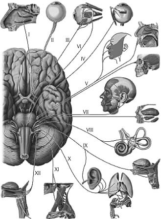
Rice. 69. Control of organs by cranial nerves, diagram. I – olfactory nerve; II – optic nerve; III – oculomotor nerve; IV – trochlear nerve; V – trigeminal nerve; VI – abducens nerve; VII – facial nerve; VIII – vestibulocochlear nerve; IX – glossopharyngeal nerve; X – vagus nerve; XI – accessory nerve; XII – hypoglossal nerve
The structure of the cerebral cortex. The cerebral cortex is formed by gray matter, which lies along the periphery (on the surface) of the cerebral hemispheres. The thickness of the cortex of different parts of the hemispheres ranges from 1.3 to 5 mm. For the first time, Kyiv scientist V.A. Betz showed that the structure and relative position of neurons are not the same in different parts of the cortex, which determines the neurocytoarchitecture of the cortex. Cells of more or less the same structure are arranged in the form of separate layers (plates). In the neocortex, most neurons form six laminae. In different sections, their thickness, the nature of the boundaries, the size of the cells, their number, etc. vary.
Outside is the first molecular plate, in which small multipolar associative neurons and many fibers of the processes of neurons in the underlying layers lie. Second outer granular plate formed by many small multipolar neurons. The third, the widest, pyramidal plate contains pyramidal-shaped neurons, the bodies of which increase in the direction from top to bottom. Fourth internal granular plate formed by small stellate-shaped neurons. In the fifth internal pyramidal plate, which is most well developed in the precentral gyrus, contains very large (up to 125 µm) pyramidal cells, discovered by V.A. Betz in 1874. The sixth multiform plate contains neurons of various shapes and sizes.
The number of neurons in the cortex reaches 10–14 billion. In addition to nerve cells, each cell plate contains nerve fibers. K. Brodman in 1903–1909 identified 52 cytoarchitectonic fields in the cortex. O. Vogt and C. Vogt(1919–1920), taking into account the fiber structure, described 150 myeloarchitectonic areas in the cerebral cortex.
Localization of functions in the cerebral cortex. In the cerebral cortex, all stimuli that come from the external and internal environment are analyzed.
In the cortex postcentral gyrus and superior parietal lobule lie nuclei of the cortical analyzer of proprioceptive and general sensitivity(temperature, pain, tactile) of the opposite half of the body. In this case, the cortical ends of the sensitivity analyzer of the lower extremities and lower parts of the body are located closer to the longitudinal fissure of the brain, and the receptor fields of the upper parts of the body and head are projected lowest at the lateral sulcus ( rice. 70A). Motor analyzer core is located mainly in precentral gyrus And paracentral lobule on the medial surface of the hemisphere (“motor cortex”). In the upper parts of the precentral gyrus and paracentral lobule, the motor centers of the muscles of the lower extremities and the lowermost parts of the body are located. In the lower part, near the lateral groove, there are centers that regulate the activity of the muscles of the face and head ( rice. 70B). The motor areas of each hemisphere are connected to the skeletal muscles of the opposite side of the body. The muscles of the limbs are connected in isolation to one of the hemispheres; the muscles of the trunk, larynx and pharynx are connected to the motor areas of both hemispheres. In both centers described, the size of the projection zones of various organs depends not on their size, but on their functional significance. Thus, the zones of the hand in the cerebral hemisphere cortex are much larger than the zones of the trunk and lower limbs combined.
On the surface of the middle part of the temporal gyrus facing the insula there is core of the auditory analyzer. Conducting pathways from the hearing organ receptors on both the left and right sides approach each hemisphere.
Visual analyzer core located on the medial surface of the occipital lobe of the cerebral hemisphere on both sides (“along the banks”) of the calcarine groove. The nucleus of the visual analyzer of the right hemisphere is connected by pathways with the lateral half of the retina of the right eye and the medial half of the retina of the left eye; left with the lateral half of the retina of the left eye and the medial half of the retina of the right eye.

Rice. 70. Location of cortical centers. A – Cortical center of general sensitivity (sensitive “homunculus”) (from V. Penfield and I. Rasmussen). Images of a cross section of the brain (at the level of the postcentral gyrus) and associated symbols show the spatial representation of the body surface in the cerebral cortex. B – Motor area of the cortex (motor “homunculus”; (from V. Pentfield and I. Rasmussen). The image of the motor “homunculus” reflects the relative sizes of the areas of representation of individual parts of the body in the cortex of the precentral gyrus of the cerebrum
Cortical end of the olfactory analyzer - it is a hook and also old and ancient bark. The old cortex is located in the area of the hippocampus and the dentate gyrus, the ancient cortex is located in the area of the anterior perforated space, the septum pellucidum and the olfactory gyrus. Due to the close location of the nuclei of the olfactory and gustatory analyzers, the senses of smell and taste are closely related. The nuclei of the taste and olfactory analyzers of both hemispheres are connected by pathways with receptors on both the left and right sides.
The described cortical ends of the analyzers carry out the analysis and synthesis of signals coming from the external and internal environment of the body, components first signaling system reality (I.P. Pavlov). Unlike the first one, second signaling system exists only in humans and is closely related to the development of articulate speech.
Speech and thinking in humans are carried out with the participation of the entire cerebral cortex. At the same time, there are zones in the cortex that are the centers of a number of special functions related to speech. Motor analyzers of oral and written speech are located in areas of the frontal lobe cortex adjacent to the precentral gyrus near the nucleus of the motor analyzer. Analyzers for visual and auditory speech perception are located near the cores of the visual and hearing analyzers. In this case, speech analyzers in right-handers are localized only in the left hemisphere, and in left-handers only in the right.
Basal (subcortical central) nuclei and white matter telencephalon. In the thickness of the white matter of each cerebral hemisphere there are accumulations of gray matter, forming separately lying nuclei, which lie closer to the base of the brain. These kernels are called basal(subcortical central). These include striatum, fence And almond-shaped body. The nuclei of the striatum form the striopallidal system, which, in turn, belongs to the extrapyramidal system involved in the control of movements and the regulation of muscle tone.
To the white matter of the hemisphere include the internal capsule and fibers passing through the cerebral commissures (corpus callosum, anterior commissure, fornix commissure) and heading to the cortex and basal ganglia; the fornix, as well as systems of fibers connecting areas of the cortex and subcortical centers within one half of the brain (hemisphere).
Lateral ventricle. The cavities of the cerebral hemispheres are lateral ventricles(I and II), located in the thickness of the white matter under the corpus callosum. Each ventricle consists of four parts: the anterior horn lies in the frontal lobe, the central part in the parietal lobe, the posterior horn in the occipital lobe, and the inferior horn in the temporal lobe.
diencephalon, located under the corpus callosum, consists of the thalamus, epithalamus, metathalamus and hypothalamus. Thalamus(visual thalamus) paired, formed mainly by gray matter, is the subcortical center of all types of sensitivity. The medial surface of the right and left thalamus, facing each other, forms the lateral walls of the cavity of the diencephalon of the third ventricle. Epithalamus includes the pineal body (epiphysis), leashes and triangles of leashes. The pineal body, which is an endocrine gland, is suspended, as it were, on two leashes connected to each other soldering and connected to the thalamus through leash triangles. The triangles of the leashes contain nuclei related to the olfactory analyzer. Metathalamus formed by paired medial and lateral geniculate bodies lying behind each thalamus. Medial geniculate body along with the lower colliculi of the plate of the roof of the midbrain (quadrigeminal) - subcortical center of the auditory analyzer. Lateral geniculate body together with the superior colliculi of the plate of the roof of the midbrain is subcortical center of the visual analyzer. The nuclei of the geniculate bodies are connected with the cortical centers of the visual and auditory analyzers.
Hypothalamus located anterior to the cerebral peduncles and includes a number of structures: located anteriorly visual part(optic chiasm, optic tract, gray tubercle, infundibulum, neurohypophysis) and olfactory part(mastoid bodies and the subthalamic region itself, the subthalamus). The functional role of the hypothalamus is very large (see section “Endocrine glands”, p. XX). It contains the centers of the autonomic part of the nervous system. The medial hypothalamus contains neurons that perceive all changes occurring in the blood and cerebrospinal fluid (temperature, composition, hormone content, etc.). The medial hypothalamus is also connected to the lateral hypothalamus. The latter does not have nuclei, but has bilateral connections with the overlying and underlying parts of the brain. The medial hypothalamus is a link between the nervous and endocrine systems. IN last years Enkephalins and endorphins, which have a morphine-like effect, are isolated from the hypothalamus. They are involved in the regulation of behavior and vegetative processes. The hypothalamus regulates all body functions except heart rate, blood pressure and spontaneous respiratory movements, which are regulated by the medulla oblongata.
Mastoid bodies, formed by gray matter covered with a thin layer of white, are the subcortical centers of the olfactory analyzer. Located anterior to the mastoid bodies gray bump, which contains the nuclei of the autonomic nervous system. They also influence a person's emotional reactions. The part of the diencephalon located below the thalamus and separated from it by the hypothalamic groove makes up the hypothalamus itself. The coverings of the cerebral peduncles continue here, the red nuclei and the black substance of the midbrain end here.
Cavity of the diencephalon - III ventricle- is a narrow, slit-like space located in the sagittal plane, limited laterally by the medial surfaces of the thalamus, below by the hypothalamus, above by the fornix, above which the corpus callosum is located. The cavity of the third ventricle passes posteriorly into the midbrain aqueduct, and in front on the sides through the interventricular foramina communicates with the lateral ventricles.
TO midbrain include the cerebral peduncles and the roof of the midbrain. Legs brain - these are white round (rather thick) cords emerging from the pons and heading forward to the cerebral hemispheres. Each leg consists of a tire and a base, the boundary between them is black matter(color depends on the abundance of melanin in its nerve cells), relating to the extrapyramidal system, which is involved in maintaining muscle tone and automatically regulates muscle function. Base of the leg formed by nerve fibers running from the cerebral cortex to the spinal and medulla oblongata and the pons. Tegmentum of the cerebral peduncles contains mainly ascending fibers heading to the thalamus, among which the nuclei lie. The largest are red kernels, from which the motor red nucleus-spinal tract begins. In addition, the tire contains reticular formation and the nucleus of the dorsal longitudinal fasciculus (intermediate nucleus).
IN roof of the midbrain differentiate roof plate(quadrigeminal), consisting of four whitish hillocks, two upper (subcortical centers of the visual analyzer) and two lower (subcortical centers of the auditory analyzer). The pineal body lies in the depression between the superior colliculi. The quadrigeminal region is a reflex center for various types of movements that arise mainly under the influence of visual and auditory stimuli. From the nuclei of these hillocks a conducting path originates, ending on the cells of the anterior horns of the spinal cord.
Midbrain plumbing(Aqueduct of Sylvius) is a narrow canal (2 cm long) that connects the III and IV ventricles. Around the water supply is located central gray matter, which contains the reticular formation, nuclei of the III and IV pairs of cranial nerves and other nuclei.
TO hindbrain include the pons, located ventrally, and the cerebellum lying behind the pons. Bridge(Varoliev pons), well developed in humans, looks like a lying transversely thickened ridge, from the lateral side of which extends to the right and left middle cerebellar peduncles. The posterior surface of the pons, covered by the cerebellum, participates in the formation of the rhomboid fossa, the anterior surface (adjacent to the base of the skull) borders the medulla oblongata below and the cerebral peduncles above. The pons consists of many nerve fibers that form pathways and connect the cerebral cortex with the spinal cord and the cerebellar cortex. Between the fibers lie the reticular formation, the nuclei of the V, VI, VII, VIII pairs of cranial nerves.
Cerebellum plays a major role in maintaining body balance and coordination of movements. The cerebellum is well developed in humans due to upright posture and the labor activity of the hands, especially developed cerebellar hemispheres. The cerebellum has two hemispheres and an unpaired median part - worm. The surfaces of the hemispheres and the vermis are separated by transverse parallel grooves, between which there are narrow, long layers of the cerebellum. Due to this, its surface area in an adult is on average 850 cm 2, and its mass is 120–160 g. The cerebellum consists of gray and white matter. The white matter, penetrating between the gray matter, seems to branch, forming white stripes, resembling in the median section the figure of a branching tree - the “tree of life” of the cerebellum ( see fig. 68). The cerebellar cortex consists of gray matter 1–2.5 mm thick. In addition, in the thickness of the white matter there are accumulations of gray, four pairs of nuclei. The nerve fibers connecting the cerebellum with other parts form three pairs cerebellar peduncles: inferior directed to the medulla oblongata, average to the bridge, upper to the quadrigeminal.
The cerebellar cortex has three layers: the outer molecular layer, the middle layer of piriform neurons (ganglionic) and the inner granular layer. The molecular and granular layers contain mainly small neurons. Large piriform neurons (Purkinje cells) measuring up to 40 microns, located in the middle layer in one row, are efferent neurons of the cerebellar cortex. Their axons, extending from the base of the bodies, form the initial link of the efferent pathways. They are directed to the neurons of the cerebellar nuclei, and the dendrites are located in the superficial molecular layer. The remaining neurons of the cerebellar cortex are intercalary (associative), they transmit nerve impulses to piriform neurons.
ATTENTION
All nerve impulses entering the cerebellar cortex reach the piriform neurons.
By the time of birth, the cerebellum is less developed than the telencephalon (especially the hemisphere), but in the first year of life it develops faster than other parts of the brain. A marked enlargement of the cerebellum is observed between the fifth and eleventh months of life, when the child learns to sit and walk.
Medulla is a direct continuation of the spinal cord. Its length is about 25 mm, its shape approaches a truncated cone, with the base facing upward. Front surface divided anterior median fissure, on the sides of which are located pyramids, formed by partially intersecting bundles of nerve fibers of the pyramidal pathways. The posterior surface of the medulla oblongata is divided posterior median sulcus, on the sides of it there are continuations of the posterior cords of the spinal cord, which diverge upward, turning into inferior cerebellar peduncles. The latter limit from below rhomboid fossa. The medulla oblongata is built of white and gray matter, the latter is represented by the nuclei of the IX–XII pairs of cranial nerves, olives, centers of respiration and circulation, and the reticular formation. White matter is formed by long and short fibers that make up the corresponding pathways. Centers of the medulla oblongata - blood pressure heartbeat and spontaneous breathing movements. Fibers of the pyramidal tracts connect the cerebral cortex with the nuclei of the cranial nerves and the anterior horns of the spinal cord.
Reticular formation is a collection of cells, cell clusters and nerve fibers located in the brain stem (medulla oblongata, pons and midbrain) and forming a network. The reticular formation is connected to all sense organs, motor and sensory areas of the cerebral cortex, the thalamus and hypothalamus, and the spinal cord. The reticular form regulates the level of excitability and tone of various parts of the central nervous system, including the cerebral cortex, and is involved in the regulation of consciousness, emotions, sleep and wakefulness, autonomic functions, and purposeful movements.
IV ventricle – This is the cavity of the rhombencephalon, which continues downward into the central canal of the spinal cord. The bottom of the fourth ventricle due to its shape is called rhomboid fossa. It is formed by the posterior surfaces of the medulla oblongata and the pons, the upper sides of the fossa are the upper, and the lower are the inferior cerebellar peduncles. In the thickness of the rhomboid fossa lie the nuclei of the V, VI, VII, VIII, IX, X, XI and XII pairs of cranial nerves.
From the book Marijuana: Myths and Facts by Lynn Zimmer7. Marijuana and the brain MYTHMarijuana kills brain cells. Long-term use of marijuana causes permanent damage to the structure and function of the brain, leading to memory loss, cognitive impairment, personality disorders and decline
From the book Nervous Diseases: Lecture Notes author A. A. Drozdov1. The brain and its structure The brain consists of two hemispheres, which are separated from each other by a deep groove reaching the corpus callosum. The corpus callosum is a massive layer of nerve fibers that connects both hemispheres of the brain.
From the book The Newest Victories of Medicine by Hugo GlaserChapter VI Brain and nerves Advances in brain surgery Many thousands of years ago, mankind knew about the operation of craniotomy. During excavations of ancient graves and burials in deep layers of the earth, skulls with well-healed
From the book Histology author V. Yu. Barsukov23. Nervous system. Brain The brain also contains gray and white matter, but the distribution of these two components is more complex here than in the spinal cord. Brain stem. All nuclei of the gray matter of the brainstem consist of multipolar nerve cells. On
From the book Neurology and Neurosurgery author Evgeniy Ivanovich Gusev1.4. Brain 1.4.1. Medulla Oblongata The medulla oblongata is a continuation of the spinal cord. The spinal cord passes into the medulla oblongata gradually, without a sharp boundary. The conventional boundary of the transition of the spinal cord into the medulla oblongata is the decussation
From the book Kinesitherapy of Joints and Spine author Leonid Vitalievich RudnitskyBRAIN The brain is divided into gray matter and white matter. Gray matter is a collection of nerve cells that is located in the cerebral cortex. Each area of the cortex is a nerve center that controls a particular function
From the book Homeopathic treatment of cats and dogs by Don Hamilton From the book Spinal Hernia. Non-surgical treatment and prevention author Alexey Viktorovich SadovBrain The brain is divided into gray and white matter. Gray matter is a collection of nerve cells that is located in the cerebral cortex. Each section of the cortex is a nerve center that controls one or another function of the body. From nerve
From the book Alcoholism author Alexander Vitalievich MelnikovBrain Damage to the brain drinking people is determined by two factors: 1) alcohol has a neurotoxic effect, that is, it directly causes the death of cells in the cerebral cortex; 2) disruption of brain functions is caused by a lack of
From the book Healthy to Death. The result of the study of the main ideas about healthy way life author AJ JacobsChapter 11 Brain Goal: become smarter There has never been a better time for fools in history. Never before have so many people believed that with hard work and the right techniques, you can improve your brain and become smarter. For decades, it was believed that intelligence was given by nature,
From the book Five Steps to Immortality author Boris Vasilievich BolotovBrain Double vision, speech retardation, impaired motor coordination, epilepsy, parkinsonism, multiple sclerosis, schizophrenia, mottled skin coloration. Source plant material: peony, cocklebur (non-rebe), mandrake, poppy, hemp, tobacco,
From the book A Healthy Man in Your Home author Elena Yurievna ZigalovaBrain The brain is located in the cranial cavity. The brain mass does not exceed 2% of the total body mass. On average, the brain of an adult male weighs 1375–1400 g. Moreover, the relative weight of the brain of men is less than that of women. So, in men, per 1 kg of body weight
BRAIN
The brain - encephalon - is the head part of the central part of the nervous system, located in the cranial cavity. Its development, like the spinal cord, occurred by closing the edges of the neural groove to form the neural tube. High concentration nerve tissue around five vesicles at the anterior end of the body led to the development of structures that varied in structure and function (diagram).
Brain structure diagram
Thus, the telencephalon is the richest in complex associative structures and is closely connected with the sense organs of the most ancient neuroepithelial type. The telencephalon is a sensory-associative area, devoid of its own motor centers. It is characterized by the development of two structures: 1) the 6a-sal ganglia in the form of striatum - an important associative center of strong, stable nervous connections such as instincts and 2) the cerebral cortex - a vast field of higher centers, rich in the ability to establish labile associations of a conditioned reflex nature.
The midbrain is dual in origin. In addition to the sensitive field, motor centers appear in it - the nuclei of the III and IV pairs of cranial nerves and the red nucleus.
The rhombencephalon has both sections - sensory and motor. Its medulla oblongata is a typical trunk brain of a “prevertebral” organism, containing visceral (receptor and effector) and somatoreceptor components (hearing, balance, etc.).
The brain is divided from the dorsal surface by a transverse fissure - fissura trausversa cerebri into the cerebrum and the rhomboid brain.
Large brain - cerebrum. It consists of the telencephalon, diencephalon and midbrain, and has two hemispheres representing the telencephalon. Right and left hemisphere cerebri - hemispheria cerebri dextrum et sinistrum are delimited dorsally by a deep longitudinal fissure - fissura longitudinalis cerebri. The hemispheres are covered from the dorsal surface by the diencephalon and midbrain. From the basal side you can see parts of the diencephalon - the pituitary gland and the optic chiasm, as well as the midbrain - the cerebral peduncle.
The rhombencephalon is the rhombencephalon. Consists of the hindbrain, which includes the cerebellum and medullary pons, and the medulla oblongata. The cerebellum is located dorsal to the medulla oblongata and behind the cerebral hemispheres. At the anterior end of the medulla oblongata on the ventral surface lies the brain bridge - pons. Medulla oblongata - medulla oblongata, s. myelencephalon directly continues into the spinal cord.
In cattle the large brain is comparatively short, wide and tall; the hemispheres are narrowed in front and greatly expanded in the back, which gives the brain a pear-shaped shape (Fig. 162).
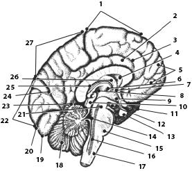
In horses, the large brain is relatively long, more laterally compressed and lower than in ruminants. There are more convolutions than in cattle (Fig. 163).
In pigs, the lateral olfactory tracts are highly developed, and the arcuate grooves are not as distinct as in the dog.
In dogs general shape the shape of the brain depends on the outline of the skull: it is sometimes more pear-shaped, sometimes more round. Three arched grooves on the mantle are typical for the dog's brain.
The brain weight of newborns differs from the brain weight of adults; it varies among different animals (Table 6).
Guinea pigs, horses, cattle, sheep are born “mature”, are born with a brain mass that is 40-60% of the brain mass of adults, while dogs, cats, rabbits, rats and mice are animals that are unable to immediately after birth, to an active existence (to independent movement, feeding), they have a relatively small brain size - 13-17% of the brain mass of adult animals. The most immature (12%) in this series is the child’s brain.
Brain development. In the early stages of ontogeny and phylogenesis, the brain represents an expanded end of the brain tube, which is called the primary, or prechordal, medullary vesicle, since it lies in front of the notochord. Its development is functionally connected with the olfactory organ, and its olfactory function is preserved in all animals, including mammals (Fig. 164).
![]()
Rice. 162. Sagittal section of the brain of cattle
Somewhat later, behind the prechordal medullary vesicle, a secondary medullary vesicle appears at the anterior end of the brain tube (it is also called the epicordal medullary vesicle, since it lies dorsal to the notochord). The development of the secondary brain bladder is due to: a) the function of the gill apparatus and lateral line organs of aquatic animals (corresponding to the organ of balance in terrestrial animals); b) differentiation of internal organs, which, in turn, was caused by an increase in metabolic rate and increased mobility of animals and c) the emergence of a primary associative and commissural center.
With the improvement of the organ of vision, the middle cerebral vesicle, the mesencephalon, is separated from the epichordal cerebral vesicle. At this trivesicular stage, the prechordal medulla is called the forebrain - prosencephalon, and the epicordal medulla - the hindbrain, or rhombencephalon. From the dorsal surface, all three parts of the brain are quite sharply delimited from each other by transverse commissures, or commissures, of nerve fibers both in front and behind the midbrain. Subsequently, the five-vesicle stage of the brain appears. From the prechordal brain the telencephalon is formed in the form of a paired brain bladder and the diencephalon.
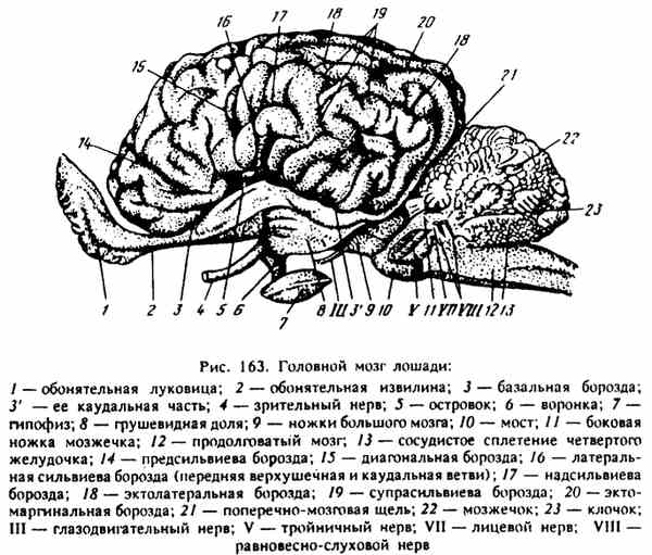
Rice. 163. Horse brain
The telencephalon reaches its highest stage of development, especially in the area of the mantle, in mammals, in particular in humans. The diencephalon in lower animals is poorly developed, but then, as the organization of animals increases, it becomes the highest subcortical sensitive center, connected with all the receptors of the analyzers and through the spinal cord with all organs of the body, as well as with the cerebral cortex as it grows. This is where the eye vesicles come from.
Almost simultaneously with the division of the prechordal brain, the rhombencephalon also differentiates into the hindbrain - metencephalon
6. Brain weight in domestic animals different types

6. Brain mass in domestic animals of different species (according to Yu.T. Tehver, 1983) and the medulla oblongata - myelencephalon. The hindbrain is initially represented by the cerebellum alone, which is the subcortical center for the correlation of muscle movements to maintain balance. Only in mammals is a medullary pons added to the cerebellum due to differentiation of the cerebral cortex. The medulla oblongata is a direct derivative of the epusordal medullary vesicle, the functions of which it retains.
Cavities of the primary brain vesicles in developed brain become cerebral ventricles. From the cavity of the prechordal brain, paired lateral ventricles arise in the telencephalon, and in the diencephalon - the third cerebral ventricle. All three ventricles are connected by the interventricular foramen (Monroe's). Due to the growth of the walls of the latter, the ventricle of the middle cerebral bladder turns into a cerebral aqueduct. The cavity of the epicordal medullary bladder becomes the fourth cerebral ventricle. It communicates with both the central spinal canal and the cerebral aqueduct.
TERMINAL BRAIN - telencephalon. It consists of two hemispheres of the cerebrum - hemispheria dextrum et sinistrum, separated from the dorsal surface by a deep longitudinal fissure - fissura longitudinalis cerebri. In each hemisphere, the mantle, olfactory brain, striatum and lateral ventricles of the brain are examined. The mantle is located on the dorsolateral surface of the hemispheres, the olfactory brain lies ventromedial. The border between the mantle and the olfactory brain on the ventral surface of the brain is the basal border, or olfactory, sulcus. The striatum lies in the ventral wall of the hemispheres, beneath the cortex but dorsal to parts of the olfactory brain.
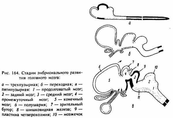
Rice. 164. Stages of embryonic brain development

Rice. 165. Cattle brain from the basal surface
The pallium is made up of gray and white brain matter. The gray brain matter - substantia grisea forms the cerebral cortex - cortex cerebri. On the dorsolateral, basal and medial surfaces it has numerous convolutions, separated from each other by deep fissures; the convolutions are covered with small grooves. Deeper fissures, convolutions and grooves have special names.
A. The grooves on the basal surface are border (Fig. 165).
1. Basal border, or olfactory, groove - sulcus rhinalis lateralis. It lies in the lateral part of the base of the brain, on the border between the olfactory brain and the mantle. The caudal part of the sulcus passes to the occipital lobe of the hemisphere, forming the occipitotemporal sulcus.
2. Medial border fissure - sulcus rhienalis medialis. Lies on the medial surface, forming the caudomedial border of the pyriform lobe.
B. Grooves on the dorsolateral surface of the mantle.
1. Lateral Sylvian fissure (fissure) - fissura sylvia. It starts from the basal border sulcus in the plane of the optic chiasm. The middle cerebral artery passes through it. It is divided into three branches: caudal, middle, or apical, and rostral. In the depths of the Sylvian fissure lies part of the cloak - the islet of Reil.
2. Ectosylvian, or first arcuate, groove - sulcus presylvius. It lies dorsal and caudal to the branches of the Sylvian fissure.
3. Suprasylvian, or second arcuate, groove - sulcus suprasylvius. It is represented by two grooves: diagonal - underdeveloped, lying above the rostral branch of the Sylvian fissure. The other, the suprasylvian fissure itself, runs parallel to the ectosylvian fissure.
4. Ectomarginal, or third arcuate, groove - sulcus ectomarginalis. Consists of two separate parts. Both run along the dorsal edge of the mantle, with the posterior portion retaining the name lateral sulcus and the anterior portion being called the coronal sulcus.
B. Grooves on the medial surface of the mantle.
1. The groove of the corpus callosum - sulcus corporis callosi runs along the dorsal edge of the corpus callosum.
2. The cingulate sulcus lies in both hemispheres above the sulcus of the corpus callosum. It is divided into the rostral part, or genu groove - sulcus genualis, and the dorsocaudal part, or ridge groove - sulcus splenialis.
Conducting pathways of the telencephalon. The white medulla - substantia alba - lies under the cortex of the cloak. It consists of pathways: associative, commissural and projection.
L. Associative, or combinational, fibers connect individual areas of the cortex within each hemisphere. They are divided into short fibers (between the gyri) and long fibers (between the lobes of the hemispheres). They pass in the outer capsule.
B. Commissural, or commissural, fibers connect areas belonging to different hemispheres. They form the corpus callosum - the largest commissure of the brain. It is placed between the hemispheres in the depth of the longitudinal slit. There is a trunk of the corpus callosum - truncus corporis callosi and two ends - anterior and posterior. The anterior end is called the knee of the corpus callosum - genu corporis callosi. It bends ventrally. The posterior end, or ridge, of the corpus callosum - splenium corporis callosi - fuses with the fornix. Commissural fibers emerging from the trunk of the corpus callosum form the radiance of the commissure - radiatio corporis callosi. Forming the roof of the lateral ventricle of the brain, it diverges into the anterior, lateral and posterior sections of the mantle cortex.
The commissural pathways pass not only through the corpus callosum, but also through four more commissures - commissures: the commissure of the visual tuberosities, or the gray commissure; the rostral commissure, located in the anterior part of the third ventricle, connects the olfactory lobes; the caudal commissure is the commissure of the vault of the lateral ventricles of the brain, located in the caudal part of the third ventricle and the commissure of the ammon's horns, located at the bottom of the lateral ventricles of the brain.
B. Projection fibers are bilateral. They connect the mantle cortex with both individual parts of the brain stem and the spinal cord. The projection fibers run in the inner capsule.
Functionally, projection pathways are divided into efferent and afferent.
Efferent pathways (also known as centrifugal, centrifugal, motor) carry impulses from the cerebral cortex to different parts of the cerebrum, rhomboid and spinal cord.
Afferent pathways (also known as centropetal, centripetal, sensitive) bring impulses to the cerebral cortex from the visual thalamus of the diencephalon.
In contrast to the cerebral cortex, the entire gray medulla of the remaining parts of the central nervous system is united by the concept of “subcortex”. Impulses from all parts of the body first flow into different sections of the subcortex (including the visual thalamus), and from the latter they already enter the cerebral cortex.
The lobes are examined on the cloak: frontal, temporal, parietal, occipital and olfactory. In front, the frontal lobe - lobus frontalis - is psychomotor, only in the dog is it clearly delimited by the coronary groove - sulcus coronarius. The occipital lobe - lobus occipitalis - visual, occupies the caudal part of the mantle behind the plane drawn through the splenium of the corpus callosum. The parietal lobe - lobus parietalis - is psychosensory, lies between the frontal and occipital lobes. The temporal lobe - lobus temporalos - auditory, is located approximately behind the lateral fissure (Sylvian) in the ventral half of the mantle. The olfactory lobe - lobus olfactorius forms the olfactory brain.
The olfactory brain - rhinencephalon - is located in the ventromedial part of each cerebral hemisphere. Its individual parts are visible on the basal and medial surfaces of the hemispheres, as well as on the bottom of the lateral ventricles of the brain. The olfactory bulbs, olfactory tracts and convolutions, olfactory triangles and pyriform lobes are concentrated on the basal surface of the hemispheres. On the medial surfaces of the hemispheres, the perioolfactory field, hippocampal convolutions, cingulate convolutions and the cut surface of the rostral commissure are visible, and on the bottom of the lateral ventricles of the brain - the caudate nuclei, the horns of Ammon (hippocampus) and the fornix.
1. Olfactory bulb - bulbus olfactorius - a paired formation in the form of a rather flat, elongated and dorsally curved hollow medullary process, which protrudes beyond the anterior edge of the cerebral hemisphere into the olfactory fossa of the ethmoid bone. The dorsomedial part of the bulb is built of gray medulla, and the lateroventral part is made of white medulla. The bulb contains the ventricle of the olfactory bulb - ventriculus bulbi olfactorii. It is a continuation of the lateral ventricle of the brain.
The olfactory bulb includes the olfactory nerves - pp. olfactorii (I pair). They contain numerous bundles of nerve fibers - fila olfactoria, directed from the olfactory cells of the nasal mucosa to the nerve cells of the bulb. Thus, the olfactory bulbs are the primary olfactory centers.
2. Olfactory pathways begin from the nerve cells of the olfactory bulb. They form the white medulla of the bulb itself and the olfactory tracts - common, medial and lateral. The lateral olfactory tract passes into the pyriform lobe, delimiting along its path the lateral olfactory gyrus - gyrus olfactorius lateralis. The medial olfactory tract reaches the medial surface of the cloak, forming the paraolfactory field - area parolfactoria. Along the way, it forms the medial olfactory gyrus - gyrus olfactorius. The olfactory tracts are limited by the olfactory triangles - trigonum olfactorium from the gray medulla. They conduct impulses from the olfactory bulb to the cells of the secondary olfactory centers located in the olfactory gyri, olfactory triangles, peri-olfactory fields, and also in the pyriform lobes.
3. The piriform lobe - lobus piriformis (hook - uncus) is located caudally from the lateral olfactory tract and the olfactory triangle, and medially borders the cerebral peduncles. The caudomedial border of the pyriform lobe is the medial border fissure, or hippocampal fissure - fissura hippocampi. The pyriform lobe contains a cavity that represents the posterior part of the lateral ventricle of the brain. The end of Ammon's horn lies on the inner wall of the lobe. The piriform lobe caudally passes without a clear boundary into the hippocampal gyrus - gyrus hippocampi, located on the medial surface of the hemisphere, posterior and lateral to the hippocampal fissure. The hippocampal gyrus continues dorsally into the cingulate gyrus - gyrus cinguli. The latter passes directly dorsally from the corpus callosum and, bending around it in front, connects with the perioolfactory field.
The piriformis lobe and the hippocampal gyrus serve as secondary olfactory centers, and the hippocampal gyrus also acts as a taste center.
4. Ammon's horn (hippocampus) - the hippocampus with its dorsal section forms the bottom of the lateral ventricle, lies behind the caudate nucleus, from which it is separated by the choroid plexus of the lateral ventricle.
The horn of Ammon is a fold of the cerebral cortex in the region of the hippocampal fissure and piriform lobe. It curves crescent-shaped laterocaudally and ventrally and rests its end against the wall of the pyriform lobe.
The horns of Ammon lie dorsally on the visual tuberosities, being separated from them by the choroid plexus of the third cerebral ventricle. Being the highest association subcortical olfactory centers, the Ammon's horns are connected with various parts of the cerebral cortex and subcortical nuclei. Their conducting paths form the fornix and its derivatives.
5. Fornix - fornix contains pathways connecting the horns of Ammon with the mastoid body of the diencephalon. Individual sections of this bundle of conducting paths are assigned various names- grooved leaves, border of Ammon's horn, legs, pillars and body of the arch and commissure of Ammon's horns.
The grooved leaf - alveus hippocampi covers the ammon's horn from its surface facing the lateral ventricle of the brain. It is built from nerve fibers originating from the gray medulla of the piriform lobe and the cornu of Ammon. Along the dorsolateral margin, these fibers create the border of the Ammon's horn, which continues into the crus of the fornix. The latter, connecting with the leg of the other side, becomes the short body of the arch. The body of the fornix serves as the roof of the third cerebral ventricle. Rostrally it divides into two columns of the vault. The latter go medially from the caudate nuclei to the mastoid body and the gray tubercle of the diencephalon (hypothalamus). The commissure of ammon's horns - commissura hippocampi is formed by transverse fibers between the legs of the arch; it connects the dorsal ends of the ammon's horns to each other.
6. Between the anterior end of the corpus callosum (beak) and the columns of the fornix lies the anterior commissure of the brain - commissura rostralis.

Rice. 166. Section of the striatum
Rice. 167. View of the lateral ventricle of the brain
![]()
Rice. 168. Stem and subcortical parts of the brain
On the border between the fence, the shell and the ammon's horn is the amygdala - corpus amygdaloideum.
The striatum is connected by conducting pathways: 1) with the cerebral cortex; 2) with the diencephalon (with the visual thalamus and hypothalamus); 3) with the nuclei of the reticular formation of the midbrain (red nucleus, etc.); 4) with the nuclei of the pons and medulla oblongata (caudal olives); 5) with the nuclei of the cranial nerves.
Various reflex circuits are closed through the striatum: a) peripheral receptor apparatus - visual thalamus, striatum - somatic and visceral effector apparatuses, or b) cortex - striatum - somatic and visceral effector apparatuses. In mammals, the nuclei of the striatum are the most important subcortical motor centers: 1) coordinated involuntary movements (walking, running, climbing); 2) regulation of muscle tone at rest and movement; 3) unconditioned reflexes in the form of gestures (in humans), posture and facial expressions and, finally, 4) higher subcortical vegetative centers. The striatum functions as a whole, but its individual parts act in the opposite way: for example, the striatum inhibits movements, and the pallid nucleus, together with the medial nuclei of the thalamus optic, on the contrary, enhances them.
Development of the telencephalon. From the anterior and ventral walls of the telencephalon come the olfactory brain - the olfactory lobes and striatum, and from its dorsal wall - the mantle.
The development of the olfactory lobes of the brain is determined by the presence of an olfactory analyzer, which in aquatic animals plays an exceptional role in orientation in the external environment. The processes of sensitive olfactory cells end in the olfactory lobes - outgrowths of the wall of the telencephalon. The distal sections of the olfactory lobes form the primary olfactory centers - the olfactory bulbs, and the proximal sections - the ancient olfactory cortex from the gray medulla in the form of olfactory convolutions and olfactory triangles; they arise as secondary olfactory centers.
In the ventral wall of the telencephalon, dorsal to the anlage, the anlage of the magnocellular basal ganglion appears very early. This is the primary and, moreover, higher motor center. The nasal ganglion is also preserved in mammals in the form of the nucleus pallidum. Later, in terrestrial animals, additional small-celled nuclei grow, forming the shell (starting with reptiles), and in mammals there is still a caudate nucleus. Both are combined under the name "striatum". With the appearance of the secondary cloak, the striatum is penetrated by internal and external capsules from conducting pathways going to the cortex of the cloak and back.
In the evolution of the cloak, two formations are observed various functions and structure, not counting the membrane-like primitive cloak, characteristic of aquatic animals and built only from ependyma.
In terrestrial animals, the growth of the secondary cloak is caused by the introduction into it of new nerve fibers (projection) from the diencephalon, which are conductors of various analyzers - skin, visual, auditory, muscle.
The cortex of the secondary cloak in a number of animals becomes extremely complex in structure, its functions and structure are sharply differentiated, and its size increases. In large mammals, the cloak is usually dotted with convolutions and furrows. The convolutions in a series of animals are located unequally. In some cases (in carnivores, ungulates) they mainly run in arcs around the transverse Sylvian fissure; In primates and humans, the gyri form two systems - the false and parietal. Both systems are separated by the Sylvian fissure. In the third group of animals, the Sylvian fissure is absent, and the fissures run longitudinally in the anterior part of the brain and transversely in the posterior part. Due to the above, absolute homologization of convolutions between different orders of animals is extremely difficult, and in some cases impossible. Small animals have no convolutions at all (animals with a smooth brain). The largest number of gyri is found in elephants and whales; small primates have no gyri. In ontogenesis, convolutions also do not appear immediately, but in a certain sequence.

Rice. 169. Cyto- and myeloarchitecture of the cerebral cortex
Cyto- and myeloarchitecture of the cerebral cortex (Fig. 169). In the process of historical development, cytoarchitectonics and myeloarchitectonics become more complex, i.e., the structure and arrangement of cells and fibers between them in the cortex of the cloak. The cellular elements of the cortex are distributed in six layers parallel to the surface of the brain. These layers, counting from the surface deep down, are as follows: I - molecular; II - external granular; III - small pyramidal cells. Cells of layers II and III appear the latest; they are credited with a higher-order association function, characterizing higher nervous activity; IV - internal granular, primary in origin, it has a receptor function; V - large pyramidal cells and VI - spindle-shaped and polymorphic cells (performs an effector function). In the primitive olfactory cortex, of the listed layers there are I, V and VI.
The pyramidal cells of V. A. Betz reach a special development both in phylogenesis and ontogenesis. The density of nerve cells in the cortex also varies: in 1 mm3 in mammals there are up to 5-10 thousand, and in primates and humans even up to 35-50 thousand.
Based on local differences in cytoarchitectonics, the mammalian cortex is divided into areas - areas. Individual fields, based on dissimilarities in myeloarchitecture, can in turn be subdivided into smaller regions. Each field is characterized by a specific function. There are fields with a function found only in humans. Naturally, such fields are absent in lower animals. Finally, there are also fields whose function is still not clear enough. The total number of fields in a person exceeds 250.
Phylogenetically, all fields are differentiated from the primitive four brain regions of lower animals (marsupials and insectivores). These areas represent the brain sections of the analyzers: the anterior lobes of the cortex are the motor analyzer, the occipital lobes are the visual ones, and the intermediate lobe is the skin analyzer. In higher animals, new association areas arise: the frontal lobe, and then the temporoparietal, with the largest volume in humans. Thus, the frontal lobes in a rabbit make up 2%, in a cat 3, in a dog 7, in monkeys 8-16, in humans 29% of the total mass of the brain.
The absolute mass of the brain varies widely, and its relative mass is inversely proportional to the mass of the animal. The absolute mass of the brain in whales is 4600-7000 g, and the relative mass is 1/10,000-1/14,000; for elephants, 4300-5400 g and 1/375-1/560, respectively; in a horse 372-570 g and 1/480-1/1000; in cattle 410-550 g and 1/600-1/770; in pigs 96-145 g and 1/1200-1/1900; in dogs 46-138 g and 1/30-1/400; in the dark tenacious monkey 126 g and 1/15; in humans 1350-1450 g and 1/35-1/45. In young animals, the relative brain mass is significantly higher than in adults: in a 5-week-old lion cub it is 1/18, and in an adult lion it is 1/546; in a newborn child it is 1/8, and in an adult it is up to 1/35.
The percentage of gray to white brain matter in small animals is higher than in large ones: in the horse 52.1 and 47.6%; in sheep 54.9 and 45.1; in dogs 61.1 and 38.9; in boars 80.7 and 19.3; in humans 40 and 60%.
DIENABRAIN - diencephalon is located between the striatal bodies of the telencephalon (in front) and the midbrain (back). Dorsally, it is covered by the vascular tectum of the third cerebral ventricle and the horns of Ammon. It consists of three sections of different structure and function: epithalamus, thalamus and hypothalamus. Epithalamus - epithalamus is formed by the vascular covering of the third cerebral ventricle, the epiphysis and the paired node of the frenulum. Thalamus - thalamus consists of the visual hillocks, between which there is a ring-shaped third ventricle of the brain. Hypothalamus - hypothalamus consists of a visual protrusion with an end plate, a gray tubercle with a funnel and an appendage of the brain - the pituitary gland and the mastoid body. All parts of the hypothalamus are visible on the basal surface of the brain between the cerebral peduncles, behind the optic chiasm.
The thalamus, or thalamus, is a paired formation, the most massive part of the diencephalon. Rostrolaterally they merge with the caudate nuclei of the striatum; the visual tuberosities are separated from the latter by a border strip, from the quadrigeminal tract by a transverse groove, and from each other by a fossa of the visual tuberosities, covered by the vascular covering of the third cerebral ventricle. The tubercles are built from an accumulation of numerous nuclei of the gray medulla. The largest of them are the following:
1) the anterior nucleus lies in the thickness of the anterior tubercle, in the rostromedial part of the optic tubercle; it is the center for switching olfactory and gustatory afferent pathways to reflex pathways;
2) the caudal nucleus is enclosed in the thickness of the caudolateral section of the lateral tubercle and is built from intermediate visual and auditory centers. From the optic chiasm on the basal surface of the brain, the visual tracts - tractus opticus - depart. Each tract bends laterally around the thalamus and passes into the lateral geniculate body - corpus geniculatum laterale, which is lost in the caudal nucleus of the optic thalamus. The geniculate body itself is the switching center for visual pathways going to the cortex. Between the lateral geniculate body and the quadrigeminal protrudes the medial geniculate body - corpus geniculatum mediale. It connects the caudal (auditory) colliculi with the caudal nucleus of the visual thalamus and serves as an intermediate auditory center on the way to the cortex;
3) the lateral nucleus consists of switching centers for the pathways of the skin analyzer going to the cortex;
4) the medial nucleus is an intermediate motor center for the extrapyramidal system;
5) the mesh formation is located between the nuclei, it contains vegetative centers.
The third ventricle of the brain - ventriculus tertius lies between the visual hillocks, has a ring-shaped shape, since the intermediate fusion of the visual hillocks grows into it. In the walls of the ventricle is the central gray matter of the third ventricle of the brain - substantia grisea centralis. It houses the subcortical autonomic centers. The third ventricle of the brain communicates caudally with the cerebral aqueduct of the midbrain, and rostrally behind the nasal commissure of the brain - commissura rostralis with the lateral ventricles of the telencephalon through the interventricular foramen.
Epithalamus. Along the edges of the fossa of the visual tuberosities, the brain stripes of the visual tuberosities are visible - striae medullares, and on them is a paired frenulum - ganglion habenulae. The knot turns into a bridle, or leash, habenula. The pear-shaped epiphysis (pineal gland) - epiphysis - is attached to it. This is an endocrine gland. It lies in the fossa between the visual tuberosities and the quadrigeminal ridge and is a time sensor. The frenulum ganglion serves as an intermediate center for reflex pathways between the brain, the nuclei of the V pair and the interpeduncular nucleus.
The vascular covering of the third ventricle - tela choroidea ventriculi tertii - is formed by a fold of the epithelial lamina of the pia mater of the brain and the choroid plexus. The epithelial plates of the tegmentum are attached along the edge of the fossa of the visual tuberosities and fornix. The vascular tegmentum separates the optic tuberosities from the ammonian horns and from the fornix; it penetrates through the interventricular foramen into the lateral ventricles of the brain in the form of the choroid plexus of the lateral ventricles of the brain - plexus choroideum ventriculi lateralis. The vascular tegmentum forms a protrusion in front of the epiphysis and directly behind the splenium of the corpus callosum.
Subthalamic region - hypothalamus. It is located ventral to the third cerebral ventricle of the brain, and the gray tubercle - tuber cinereum lies directly behind the optic chiasm, between the peduncles of the cerebrum. It is the autonomic center and connects to the visual thalamus and the olfactory brain. In the center of the gray tubercle there is a funnel bay (protrusion of the ventral wall of the ventricle). The funnel itself is thin-walled infundibulum; The pituitary gland is attached to it. The appendage of the brain, or pituitary gland, the hypophysis cerebri, is a flat-round body of complex structure with a small central cavity. The pituitary gland consists of three parts: medullary (cranial), intermediate and glandular (caudal). It, being a powerful endocrine gland that secretes various hormones, acts as a vegetative center.
The mastoid body - corpus mamillare lies directly behind the gray tubercle and serves as an intermediate reflex olfactory center, which connects to the olfactory brain through a complex of formations of the fornix. In addition, the mastoid body is associated with the visual tuberosities and the reticular formation. The dog has a paired mastoid body.
Circumtubercle - metathalamus. On the posterolateral part of the visual thalamus there is a cushion - pulvinar, which turns into two small elevations - the lateral and medial geniculate bodies - corpus geniculatum laterale et mediale. The lateral geniculate body passes ventrally into the optic tract, and the medial geniculate body into the anterior legs of the quadrigeminal midbrain, from which it is separated by a transverse groove. At the base of the cushion and the lateral geniculate body is the caudal optic nucleus. The geniculate body itself is the center for switching visual pathways going to the cerebral cortex. The caudal geniculate bodies of both sides of the visual thalamus connect the caudal (auditory) colliculi with the caudal nuclei of the visual thalamus and are intermediate auditory centers on the way to the cortex.
Development of the diencephalon. The diencephalon in the primitive form is formed from a small number of cells in the wall of the extensive third ventricle of the brain; only in terrestrial animals, and especially in mammals, does it reach a significant size.
1. The well-defined embryonic tegmental plate in all adult mammals forms the epithalamus. The pineal gland is preserved as a rudiment of the third unpaired parietal eye. Only some aquatic animals and reptiles have an eye-shaped bladder under the skin. In mammals, the pineal gland becomes an endocrine gland, the function of which has not yet been sufficiently studied.
2. A unique and highly developed embryonic floor plate forms the subthalamic part of the diencephalon - hypothalamus.
The pituitary gland comes from three different sources. Thus, the ectoderm of the pharynx (Rathke’s pouch) turns into a branched gland. The lumen of the gland subsequently disappears, but strands of glandular cells of various structures remain, surrounded by a large number of vessels. In terrestrial animals, the nervous part of the pituitary gland arises from the wall of the funnel from nerve (glial) cells and nerve fibers. And finally, the intermediate part of the pituitary gland is separated from epithelial cells. The pituitary gland as a whole secretes over a dozen different hormones, which from the glandular part enter directly into the blood, and from the nervous and intermediate parts into the cerebrospinal fluid. The pituitary gland interacts with the autonomic centers located in the walls of the third ventricle of the brain.
3. The embryonic lateral plate gives rise to the visual tubercles - thalami. The thalamus includes the nuclei of the visual thalamus.

Rice. 170. Midbrain in cross section
They serve as: 1) intermediate centers of all conducting pathways, which are directed to the cerebral cortex and conduct various impulses - olfactory, skin and muscle sensitivity, taste, and in terrestrial animals, in addition, visual and statoacoustic; 2) the intermediate center of all pathways coming from the cape cortex to various parts of the brain. This explains why the visual thalamus begins to form with the appearance of the olfactory cloak (in reptiles) and reaches its maximum development in mammals due to the formation of the secondary cloak. The powerful evolution of the visual thalamus is also facilitated by the movement of visual centers from the midbrain to the intermediate brain and, finally, communication with the cerebellum. As a result of the enrichment of the thalamus with intermediate centers, the intermediate mass of the visual thalamus increases, which, penetrating into the cavity of the third ventricle of the brain, turns it into a ring-shaped canal. IN gray matter The walls of the third cerebral ventricle contain numerous higher subcortical autonomic centers.
MIDDLE BRAIN - mesencephalon consists (Fig. 170): of the legs of the cerebrum; quadrigeminal plates and tires or caps. The midbrain cavity has turned into the cerebral aqueduct - aqueductus mesencephali; it connects the third and fourth cerebral ventricles. The walls of the aqueduct contain the central gray medulla.
The peduncles of the large brain - pedunculi cerebri - protrude on the basal surface of the brain in the form of two thick ridges between the optic tracts and the brain bridge. They are separated by an interpeduncular groove. The third pair of cranial nerves - the oculomotor nerve - n. oculomotorius is separated from the legs. All pathways connecting the cortex and visual thalamus with the middle, rhomboid and spinal cord pass through the legs. Therefore, the legs are larger in those animals that have a more powerful cerebral cortex.
The roof plate (quadrigeminal) - lamina tecti represents the dorsal part of the midbrain; it lies caudal to the optic thalamus and nasal to the cerebellum. The plate consists of paired rostral and caudal hills - colliculi rostrales et caudales.
The hills are separated by transverse and median furrows. On the surface, the quadrigeminal plate is covered with white medulla, under which lies gray medulla; in the rostral hills it is the subcortical visual center, and in the caudal hills it is the subcortical auditory center.
In general, the quadrigeminal plate is the coordinating center of a number of impulses: visual, auditory, olfactory, general sensitivity (from the medial lemniscus) and impulses from the cerebral cortex. Motor impulses are transmitted to the spinal cord, as well as to the eye muscles, the red nucleus, the cerebellum and the pons.
Between the quadrigeminal plate and the cerebral peduncles there is a covering of the legs, or cap, - tegmentum mesencephali. It contains paired nuclei of the gray medulla; in the plane of the anterior hills lie: the red nucleus - the nucleus ruber - the motor center of the spinal cord; nucleus of the oculomotor nerve - nucleus motorius n. oculomotorii (III pair); parasympathetic nuclei of the oculomotor nerve (see below); caudal are the nucleus of the trochlear nerve - nucleus motorius n. trochlearis (IV pair) and part of the nucleus of the V pair of nerves. Through the entire cap, from the medulla oblongata to the intermediate brain, a reticular formation passes through - formatio reticularis, forming the nuclei tegmenti, as well as pathways from the spinal cord and cerebellum to the quadrigemulus, to the optic thalamus and to the spinal cord.
Development of the midbrain. The midbrain in lower animals and in embryos of higher animals reaches significant sizes. The gray medulla of the embryonic lateral plates of the mesencephalon, growing, forms the fornix of the mesencephalon, from which in lower animals (including birds) the colliculus, or optic lobes, arises. Initially, the colliculus was the highest visual center, since the optic nerves and pathways from the striatum end there. But already in reptiles, some of the fibers, and in mammals, almost all the fibers of the optic nerve move to the visual thalamus of the diencephalon. Because of this, the optic lobes lag behind in growth, and the visual tuberosities, on the contrary, grow.
In terrestrial animals, equilibrium-auditory centers are formed in the fornix of the midbrain, initially in the form of microscopic formations, and later macroscopic ones (in some reptiles and birds). Only in mammals, instead of a colliculus, a quadrigeminal structure appears - tectum mesencephali. In animals with good hearing (nocturnal predators), the posterior auditory colliculi predominate, while in animals with good vision, the anterior visual colliculus predominates, for example in domestic ungulates.
The cap is formed from the embryonic main plate in the ventral wall of the midbrain vesicle. As a result of the formation of the cap vault, the cavity of the cerebral bladder turns into the cerebral aqueduct. The cap contains the nuclei of the III and IV pairs of cranial nerves and special motor nuclei of the cap. The latter include the red nucleus, which connects the cerebellum with the spinal cord, and the intercortical nucleus, which connects through the frenulum ganglion to the olfactory brain.
In connection with the development of the secondary cloak in mammals, the cap is ventrally covered with a layer of white medulla from pathways coming from the cerebral cortex and visual thalamus to the rhomboid and spinal cord, and afferent pathways going to the cerebral cortex. These pathways form the cerebral peduncles, the severity of which corresponds to the degree of development of the cerebral cortex.
The rhombencephalon is divided into the medulla oblongata and the hindbrain. The hindbrain consists of the cerebellum and the medullary pons. Between the cerebellum and the medulla oblongata is the fourth cerebral ventricle.
The medulla oblongata - medulla oblongata continues caudally without a noticeable transition to the spinal cord. On its basal surface the ventral median groove is clearly visible - fissura mediana ventralis. On both sides of it there are lateral grooves; caudally they join the ventral median sulcus. Between these three grooves protrude two narrow ridges - pyramids - pyrames medullae oblongatae. They contain lateral pyramidal pathways from the cerebral cortex to the spinal cord. When passing into the lateral cords of the spinal cord, they intersect from right to left and vice versa and form the decussation of the pyramids - decussatio pyramidum, which forms the apexes of the pyramids. Laterally from the base of the pyramids, the VI pair of cranial nerves emerges from the brain - the abducens nerve - n. abducens. Near the crossroads at the tops of the pyramids and laterally the XII pair - the hypoglossal nerve - n. hypoglossus is separated from them. Laterally, three more nerves depart one after another from the hypoglossal nerve: XI pair - accessory nerve - n. accessorius, directly in front of it X pair - vagus nerve - n. vagus and even more rostrally - IX pair - glossopharyngeal nerve - n. glossopharyn -geus.
The gray medulla of the medulla oblongata is grouped into separate nuclei, from which the V, VI, VII, IX, X and XII pairs of cranial nerves emerge: a) sensory and motor; b) intermediate nuclei; c) nuclei of the VIII pair and the rostral and caudal olives associated with them (motor centers). Among the nuclei lies a mesh formation - formatio reticularis of intertwined nerve fibers and nerve cells between them, which from the medulla oblongata passes into the cap of the midbrain, and into the diencephalon. It performs primarily association and coordination functions between the various nuclei of the rhomboid and midbrain and is the center of respiration and the cardiovascular system, as well as an energy generator.

Rice. 171. Diagram of the conductive nerves of the brain and spinal cord
The white medulla of the medulla oblongata contains a large number of bundles of pathways coming from the spinal cord to various parts of the brain and pathways connecting the brain to the spinal cord (Fig. 171).
In the medulla oblongata, the motor nuclei of the cap deserve special attention - nuclei tegmenti (otherwise reticular formation - formatio reticularis), which first appear in fish and are the oldest association and motor center.
In terrestrial animals, due to the motor nucleus of the cap, the rostral and caudal olives are formed as association centers. Rostral olives appear only in terrestrial animals, starting with amphibians (due to the development of their hearing organ). They serve as an intermediate center on the way from the cochlear nerve (VIII pair) to the optic thalamus. The bundle of these fibers forms a trapezoidal body. The caudal olives are formed even later - in birds and mammals. They are connected with the nuclei of the dorsal funiculi with the visual thalamus, the cerebellum and the spinal cord. By their connections, the caudal olives are closely related to maintaining balance.
Hindbrain -metencephalon. It has two parts: the medullary pons and the cerebellum.
The brain bridge - pons lies at the anterior end of the medulla oblongata, on its border with the midbrain, in the form of a transverse ridge, the ends of which bend dorsally to the cerebellum, forming the lateral cerebellar peduncles - brachia pontis. The pons and peduncles consist of pathways connecting the pontine nuclei - nuclei pontis - with the cerebellar nuclei. In the pontine nuclei, the pathways from the cerebral cortex end and the pathways in the cerebellar hemisphere begin. From the lateral sections of the bridge, the V pair - the trigeminal nerve - n. trigeminus, the most massive of all cranial nerves, is separated. It emerges from two roots: the ventral motor and dorsocaudal sensory. On the latter there is a large semilunar (trigeminal) ganglion - ganglion trigeminal. Caudal to the bridge, also in the transverse direction, lies a trapezoidal body - corpus trapezoideum in the form of a narrow and low ridge. It is formed by pathways coming from the nuclei of the auditory nerve. From the lateral parts of the trapezoid body emerge the VIII pair - vestibulocochlear nerve - n. vestibulocochlearis and VII pair - facial nerve - n. facialis.
The cerebellum - cerebellum has an almost spherical shape; It is divided by two grooves into a middle part - the worm and two lateral lobes. The gray medulla forms the cerebellar cortex - cortex cerebelli and, in addition, individual nuclei located in the centrally located white medulla of the cerebellum.
The surface of the worm's bark is indented with transverse grooves and crevices. The worm is divided into anterior, middle and posterior lobes by two main slits. Each of them is associated with the cerebellar peduncles - anterior, middle and posterior, consisting of pathways. The anterior and posterior ends of the worm are curved ventrally and towards each other; between them there remains only a small gap - the top of the tent, or the roof of the fourth ventricle - tegmentum ventriculi quarti. The white brain matter of the worm in a sagittal section resembles a thuja branch, as a result of which it received the name tree of life - arbor vitae (as the thuja was formerly called). In the white brain matter of the worm there is a tent nucleus - nucleis fastigii, which is the subcortical center of the equilibrium analyzer.
The vermis is connected to the medulla oblongata via the caudal medullary velum - velum medullare caudale, and to the quadrigemuls via the rostral medullary velum - velum medullare rostraie.
The hemispheres of the cerebellum - hemispheria cerebelli, like the vermis, are built from numerous lobules, one of which - a flocculus in the form of a small appendage - is adjacent directly behind the lateral peduncle. In the white medulla of the cerebellar hemispheres there is a dentate nucleus - nucleus dentatus, which serves as a transmission center for motor impulses.
The cerebellum is connected to the medulla oblongata by the caudal cerebellar peduncles, to the medullary pons by the lateral peduncles, and to the midbrain by the nasal peduncles.
The caudal peduncles of the cerebellum - pedunculi caudale, or rope bodies, protrude in the form of two rollers on the dorsal surface of the medulla oblongata. Conducting pathways pass through them.
The rostral peduncles of the cerebellum - pedunculi rostraie, or connecting legs, protrude under the posterior colliculi of the quadrigemina into the peduncles of the cerebellum. The rostral peduncles lie on the dorsal surface of the medulla oblongata. Conducting paths pass through them: a) from the spinal cord to the vermis; b) from the dentate nuclei of the cerebellar hemispheres to the red nucleus and c) to the nuclei of the visual thalamus. The cerebellum's own pathways are represented by fibers between the cerebellar cortex and its nuclei and association fibers connecting individual convolutions to each other in the sagittal plane.
The cerebellum develops in connection with its function of maintaining body balance and maintaining muscle tone. Therefore, it is expressed most strongly in animals that swim, run, jump or fly quickly, and weaker in animals that move slowly. In its primitive form, the cerebellum is an unpaired plate of white or gray brain matter. The cerebellum is formed in the middle part of the embryonic tegmental plate of the rhombencephalon, and the anterior and posterior medullary velum are formed from the anterior and posterior parts of the latter. The cerebellar plate, growing from front to back, bends in an arcuate manner in the dorsal direction. Thanks to the appearance of longitudinal grooves, the middle part of the plate is separated - the body of the cerebellum and paired lateral parts - the cerebellar auricles. The body of the cerebellum in terrestrial animals is divided by transverse grooves into the anterior, middle and posterior lobes, on which additional transverse grooves appear. The anterior lobe is connected to the muscles of the head, and the middle and posterior lobe are connected to the muscles of the trunk and limbs.
In mammals, the middle lobe predominates. Longitudinal grooves on it separate the middle, unpaired part - the worm - vermis. The worm contains centers for coordinated, synchronous movements of the torso and limbs. The cerebellar hemispheres are most strongly expressed in higher mammals, which have, to a greater or lesser extent, the ability of isolated movements of the limbs. The improvement of this ability, in turn, depends on the power of the cerebral cortex as the highest center nervous activity and from the emergence of connections between the cerebellum and the cerebral cortex through its lateral peduncles and the pons. Due to the above, the cerebellar hemispheres and the pons reach their maximum development in primates and especially in humans.
The homogeneous function of the cerebellum among animals explains the rather uniform histological structure of its cortex, in which a superficial molecular layer, a middle granular layer and a deep layer of large Lurkine cells are distinguished.
The ears of the primitive cerebellum in aquatic animals are related to the organs of balance, i.e., the organs of the lateral line, and to the muscles of the tail. With the reduction of these organs in terrestrial animals, the ears also become smaller. In mammals, fragments of them are preserved - floculi, connecting to the posterior lobe of the worm.

Rice. 172. Medulla oblongata from the dorsal surface
The fourth cerebral ventricle - ventriculus quartus is located between the cerebellum and the medulla oblongata (Fig. 172). Its arch is the worm and brain sails, and its bottom is the medulla oblongata and the bridge.
At the bottom of the fourth ventricle there is a rhomboid fossa - fossa rhomboidea. The median and two lateral grooves at the bottom of the fossa distinguish a paired median eminence - eminentia medialis, on which the facial hillock - colliculus facialis - is visible opposite the lateral cerebellar peduncle. In the area of the facial colliculus lie the nuclei of the abducens (VI pair) and facial (VII pair) nerves. At the caudal end of the median eminence extends the field of the hypoglossal nerve with the nucleus of the same name.
Laterally from the field of the hypoglossal nerve, the nuclei of the IX and X pairs of nerves protrude. They form a gray wing. The area of the posterior end of the gray wings is known as the “writing pen” - calamus scriptorius.
Directly behind the lateral cerebellar peduncles and medially from them, the vestibular fields - areae vestibulares - protrude in the form of small elevations. They contain the vestibular and cochlear nuclei of the VIII pair of nerves. The cochlear nuclei lie laterally.