The bark is represented by a layer gray matter 3-5 mm thick. There are up to 15 billion or more neurons in the cortex, and the number of gliocytes in the brain is more than 100 billion. Development. Formation of the crust cerebral hemispheres occurs through the regular migration of neuroblasts of the ependymal layer along vertically oriented radial gliocytes. The most superficial and deep layers of the cortex appear first. Then the next successive waves of migration of groups of neuroblasts arise, which differentiate into neurons of the Vth, then the IVth layer, etc. Thus, the neuroblasts of the next wave of migration overcome the layer of neurons that arose from the earlier wave of migration. This creates layer-by-layer (screen) cytoarchitecture of the cortex big brain. Complex relationships are established between neurons in accordance with their place in the composition reflex arcs. Nuclear and screen nerve centers are formed. Close relationships during histogenesis develop between neurons and glial cells.
Maturation of the cerebral cortex - formation of the neural organization of the cerebral cortex in the process of child development. In the development of K. b. In ontogenesis, two processes are distinguished - the growth of the cortex and the differentiation of its nervous elements. The most intensive growth in the width of the cortex and its layers occurs in the first year of life, gradually slowing down and stopping at different times - by 3 years in projection areas, by 7 years in associative areas. The growth of the cortex occurs due to the expansion of the interneuronal space (cell rarefaction) and as a result of an increase in the fibrous component - the growth and branching of dendrites and axons - and the development of glial cells, which provide metabolic support for developing nerve cells, which increase in size.
The process of neuronal differentiation, also beginning in early postnatal ontogenesis, continues over a long period of individual development, subject to both genetic factors and external environmental influences.
The concept of GNI. The role of I.M. Sechenev and I.P. Pavlova in the development of the doctrine of GNI.
Higher nervous activity- these are processes occurring in the higher parts of the central nervous system animals and humans. These processes include a set of conditioned and unconditioned reflexes, as well as higher mental functions that ensure adequate behavior of animals (including humans) in changing environmental and social conditions.
Higher nervous activity should be distinguished from the work of the central nervous system by synchronizing the work various parts organisms with each other. Higher nervous activity is associated with neurophysiological processes taking place in the cerebral cortex brain and the subcortex closest to it.
The role of Sechenev and Pavlov.
The luminaries of science K. A. Timiryazev and I. P. Pavlov called I. M. Sechenov the father of Russian physiology. In fact, no one had conducted experiments on the brain before him. Their result was the psychophysiological and philosophical work “Reflexes of the Brain” (1863). This is where research into higher nervous activity begins. Here are its main provisions.
Memory is the basis of mental development. Several types of memory identified by Sechenov - visual, tactile, auditory, muscular, associative - are studied in subsequent physiology. Let us add to this his discovery of the phenomenon of inhibition in the central nervous system, the discovery of the psychological mechanisms of thinking, and the fundamental significance of I.M. Sechenov’s works in the development of ideas about higher nervous activity will become clear.
The ideas of I.M. Sechenov had a great influence on the young I.P. Pavlov. For forty years, I.P. Pavlov developed them, along with research on pharmacology, physiology of blood circulation, and digestion, which led to a new stage in his work - the study of higher nervous activity. In 1923, his general work “Twenty years of experience in the objective study of the higher nervous activity of animals” appeared, followed by “Lectures on the work of the cerebral hemispheres” (1927) and other works.
By higher nervous activity, I.P. Pavlov understood the activity of the cerebral hemispheres with the nearby subcortex, which determine reflexes.
IP Pavlov's studies of the relationships between excitation and inhibition, their strength and duration, made it possible to identify four main types of higher nervous activity in humans.
1. Unbalanced type. With it, excitation processes predominate over inhibition processes.
2. Balanced type with high mobility of nervous processes.
3. Balanced type with low mobility of nervous processes.
4. Weak type. In such individuals, both excitation and inhibition are poorly developed.
These four types of higher nervous activity, discovered by I. P. Pavlov, correspond to the four types of human temperaments (characters). Since the time of Hippocrates, the unbalanced type with a predominance of excitations has been referred to as the choleric temperament. Balanced with great mobility of excitation and inhibition is characteristic of the sanguine character; a balanced type with little mobility of some and other processes is inherent in phlegmatic people; the weak type with weak excitation and inhibition is characteristic of people of melancholic temperament. But it should be noted that you will not meet people with an absolutely “pure” type of temperament; in specific people, one of the types only predominates.
I. P. Pavlov’s discovery of the second signaling system, inherent only to humans, laid the foundation for the development of issues of thinking, the emergence and development of speech.
Meaning of the word. According to I.P. Pavlov, a word represents a conditioned stimulus for a person, causing irritation that is perceived and processed by the cerebral cortex. Therefore, the second signaling system with its speech apparatus is the physiological basis of thinking. Words are “signals of signals,” that is, signals for the operation of the first signaling system. Thanks to the second signaling system, the social experience of generations is used, assimilated through language and speech. If for animals words are sounds and stimuli, then for humans they are concepts.
They act with their content and meaning. Words act on functions through the nervous system internal organs, due to which they (words) are used by psychotherapists for treatment functional disorders. In the same way, words can have a harmful effect on health. This implies the need for careful use of them in communication, especially when communicating between a doctor and patients. On this score, a whole direction in medicine arose - medical ethics - the doctrine of moral principles in the activities of a doctor, including the meaning of the word.
Speech appeared as a result of communication between people in the process of work. The meaning of the word is not limited to communication, but forms the basis of abstract thinking and determines people's behavior. Through speech, information is analyzed and summarized, as well as judgments and conclusions. Speech reflects intelligence.
The cortex is composed of a layer of gray matter 3-5 mm thick. There are up to 15 billion or more neurons in the cortex, and the number of gliocytes in the brain is more than 100 billion.
Development. The formation of the cerebral cortex occurs through the regular migration of neuroblasts of the ependymal layer along vertically oriented radial gliocytes. The most superficial and deep layers of the cortex appear first. Then the next successive waves of migration of groups of neuroblasts arise, which differentiate into neurons of the Vth, then the IVth layer, etc. Thus, the neuroblasts of the next wave of migration overcome the layer of neurons that arose from the earlier wave of migration. This creates a layer-by-layer (screen) cytoarchitectonics of the cerebral cortex.
Complex connections are established between neurons relationships in accordance with their place in the reflex arcs. Nuclear and screen nerve centers are formed. Close relationships during histogenesis develop between neurons and glial cells.
Structure. All cortical neurons are multipolar. Among them, pyramidal and non-pyramidal (stellate, basket-shaped, spindle-shaped, arachnid and horizontal) neurons are distinguished based on the shape of the cells. Pyramidal neurons, most characteristic of the cortex, have a body shaped like a pyramid, the apex of which faces the surface of the cortex.
From the base pyramidal cell an axon with collaterals (recurrent, horizontal, oblique) departs. Long dendrites (apical and basal) extend from the apex and lateral surfaces of the body. The apical dendrites of a group of neurons are combined into dendritic bundles. On the surface of the dendrites of one pyramidal neuron there can be up to 4-6 thousand special receptor devices - spines. The presence of the actomyosin complex in the latter makes it possible to change the area of synaptic contact and, therefore, influence the synaptic connection.
Size of pyramidal cell body varies from 10 to 150 microns. There are small, medium, large and giant pyramids. Pyramidal cells are efferent neurons of the cortex; their axon collaterals form 3/4 of all synapses in the cortex.
Stellate neurons have a star-shaped body. Dendrites extend in all directions from the body of the stellate neuron. They are in most cases short and lack spines. The axons of stellate cells form complex branches around the cell. These are the so-called pericellular web-like axon networks. These cells are found in the lower layers of the cortex.
Basket cells(small and large), located in P-m and Sh-th layers cortex, with their numerous processes form synaptic connections with the bodies of pyramidal neurons of the V-ro layer. The cells contain a transmitter (GABA), which inhibits the transmission of excitation.
Neurogliomorphic cells found in all layers of the cortex. These are small multipolar neurons with short branching dendrites and axons.
Bipolar neurons- a small group of cells from the body of which an axon and dendrite extend. In general, the ratio between pyramidal and other forms of neurons is 85:15, that is, in favor of pyramidal neurons.
Cytoarchitecture. In the motor zone of the cortex, six main layers are distinguished: molecular, external granular, pyramidal, internal granular, ganglion, layer of polymorphic cells.
In the first (outer) molecular layer almost no cell bodies of neurons. Single horizontally oriented neurons, tangential branching of nerve fibers of underlying neurons and glial cells are detected.
Second, or outer granular, the layer contains small stellate and pyramidal neurons measuring about 10 μm. The axons of these neurons end in the III, IV and VI layers of the cortex, and the dendrites rise into the molecular layer.
Third layer- This is a layer of medium and large pyramidal neurons. The axons of these cells form associative nerve fibers that run through the white matter and connect adjacent areas of the cortex.
Fourth, or internal granular, the layer contains mainly small stellate neurons. The axons of these cells branch within the neighboring layers of the cortex, both above and below. This layer is highly developed in the visual and auditory cortex. It consists of sensory stellate neurons that have numerous associative connections with neurons of other types.
Fifth - ganglionic- the layer is formed by large pyramidal neurons (Betz cells). The apical dendrites of neurons are directed into the molecular layer. The axons of these cells go into the white matter, forming commissural and projection nerve fibers, and above all the pyramidal tracts.
Sixth layer - layer of polymorphic neurons- also contains many efferent pyramidal neurons. In addition, there are spindle neurons. The dendrites of neurons in the sixth layer penetrate the entire thickness of the cortex, reaching the molecular layer.
Maturation of the cerebral cortex - the formation of the neural organization of the cerebral cortex in the process of child development. In the development of K. b. In ontogenesis, two processes are distinguished - the growth of the cortex and the differentiation of its nervous elements. The most intensive growth in the width of the cortex and its layers occurs in the first year of life, gradually slowing down and stopping at different times - by 3 years in projection areas, by 7 years in associative areas. The growth of the cortex occurs due to the expansion of the interneuronal space (cell rarefaction) and as a result of an increase in the fibrous component - the growth and branching of dendrites and axons - and the development of glia cells, which provide metabolic support to developing nerve cells, which increase in size. The process of neuronal differentiation, also beginning in early postnatal ontogenesis, continues over a long period of individual development, subject to both genetic factors and environmental influences. The first to mature are the afferent and efferent pyramids of the lower layers of the cortex, later - located in the more superficial layers. Various types of interneurons gradually differentiate. Earlier, spindle-shaped cells mature, switching afferent impulses from subcortical structures to developing pyramidal neurons. Stellate and basket cells, which ensure the interaction of neurons and the circulation of excitation within the cortex, mature later. Ending with excitatory and inhibitory synapses on the neuron bodies, these cells create the possibility of structuring the impulse activity of neurons (alternating discharges and pauses), which is the basis of the nervous code. The differentiation of interneurons, which began in the first months after birth, occurs most intensively in the period from 3 to 6 years. Their final typification in the anterior associative areas of the cortex is noted by the age of 14. Functionally important factor The formation of the neural organization of the cerebral cortex is the development of processes of nerve cells - dendrites and axons, forming a fibrous structure. The axons through which afferent impulses enter the cortex are covered with a myelin sheath during the first three months of life, which significantly accelerates the flow of information to the nerve cells of the projection cortex. Vertically oriented apical dendrites ensure the interaction of cells of different layers of the cortex. In the projection zone they mature in the first weeks of life, reaching 6 months. third layer. Growing to the surface of the layers, they form final branches. Basal dendrites, which unite neurons within one layer, have wide branches on which multiple contacts of axons of other neurons are formed. The growth of basal dendrites and their branches increases the receptive surface of nerve cells. The specialization of neurons in the process of their differentiation and the increase in the number and branching of processes create conditions for the unification of neurons of different types into cell groups - neural ensembles. Neuronal ensembles also include glial cells and vascular branches, which ensure cellular metabolism within the neuronal ensemble. Stages of formation of the ensemble organization of nerve cells of the cerebral cortex in ontogenesis. In the formation of an ensemble organization in ontogenesis, qualitatively different stages are distinguished. By the time of birth, vertically located pyramidal cells in nearby layers and their apical dendrites create the prototype of a column, which in newborns is poor in intercellular connections. 1 year of life is characterized by an increase in the size of nerve cells, differentiation of stellate interneurons, an increase in dendrites and axonal branches; an ensemble of neurons is distinguished as a structural unit surrounded by thin vascular branches. By the age of 3, the ensemble organization is complicated by the development of nest groups, including different types neurons. At 5-6 years, along with the ongoing differentiation and specialization of nerve cells, the volume of horizontally located fibers and the density of capillary networks surrounding the ensemble increases. This contributes further development interneuronal integration in certain cortical areas. By the age of 9-10 years, the structure of the processes of interneurons and pyramids becomes more complex, the diversity of ensembles increases, and wide horizontal groupings are formed, including and uniting vertical columns. At 12-14 years of age, various specialized forms of pyramidal neurons are clearly expressed in neural ensembles, high level interneurons reach differentiation; in ensembles of all areas of the cortex, including associative cortical zones, due to the branching of the processes, the specific volume of fibers becomes significantly higher than the specific volume of cellular elements. By the age of 18, the ensemble organization of the cortex reaches adult levels in its characteristics. References: ◄ Section III. Age-related psychophysiology
text_fields
text_fields
arrow_upward
The human brain develops from the embryonic ectoderm overlying the notochord. From the 11th day of intrauterine development, starting from the head end of the embryo, the formation of neural plate, which subsequently (by week 3) closes into a tube. Neural tube detaches itself from the ectodermal layer and becomes immersed underneath it. Simultaneously with the formation of the neural tube, paired strips are laid under the ectoderm layer, from which ganglion plates are formed (neural crests).
The part of the neural tube from which the hindbrain is formed is the first to close. The closure of the tube in the anterior direction occurs more slowly than in the posterior direction due to its greater thickness. The last hole to close is at the anterior end of the neural tube. The formed neural tube expands at the anterior end, where the future brain is formed.
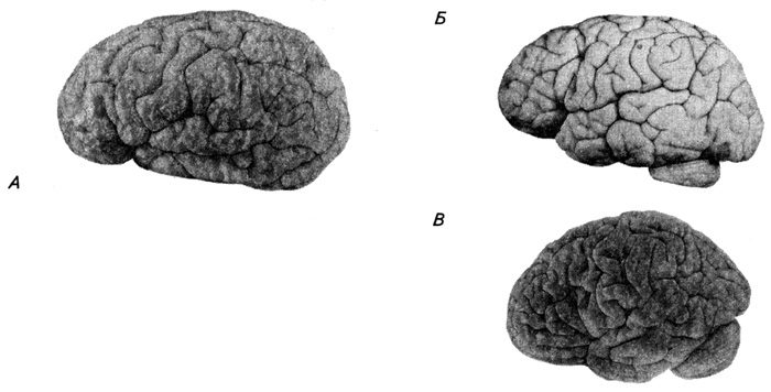
In the primary anlage of the brain, two interceptions appear and are formed three primary brain vesicles: anterior (prosencephalon), middle (mesencephalon) And posterior (rhombencephalon)(Fig. 3.49, A). In a three-week embryo, the division of the first and third bubbles into two more parts is planned, in connection with which the next one begins, pentavesical stage development (Fig. 3.49, B).
A – 3 weeks; B – 5 weeks; C – 5 months, D – 6 months; D – newborn: a – anterior, b – middle and c – posterior bladders; d – spinal cord; e – terminal, f – intermediate, g – hindbrain and h – accessory brain; 1 - optic vesicle; 2 – auditory vesicle; 3 – heart; 4 – mandibular process; 5 – olfactory tubercle; 6 – cerebral hemisphere; 7 - midbrain; 8 – cerebellum; 9 – medulla oblongata; 10 – spinal cord; 11 – larynx; 12 – inferior precentral, 13 – central, 14 – lateral, 15 – postcentral, 16 – interparietal and 17 – superior temporal sulcus; 18 – island. Roman numerals indicate cranial nerves
From the anterior bladder, a paired secondary bladder protrudes forward and to the sides - telencephalon(telencephalon), from which the cerebral hemispheres and some basal ganglia develop, and the posterior part of the anterior bladder is called diencephalon. On each side of the diencephalon, an optic vesicle grows, in the wall of which the nerve elements of the eye are formed. Develops from the posterior bladder hindbrain (metencephalon), including the cerebellum and pons, and additional (myelencephalon). The midbrain is preserved as a single whole, but during development significant changes occur in it associated with the formation of specialized reflex centers related to vision and hearing, as well as to tactile, temperature and pain sensitivity.
The primary cavity of the brain tube also changes. In the area of the telencephalon, the cavity expands into paired lateral ventricles; in the diencephalon turns into a narrow sagittal fissure - third ventricle; in the midbrain remains in the form of a canal - cerebral aqueduct; in the rhomboid vesicle it does not divide during the transition to the five-vesicle stage and turns into a common one for the hindbrain and accessory brain fourth ventricle. The brain cavities are lined with ependyma (a type of neuroglia) and filled with cerebrospinal fluid.
Due to the rapid and uneven growth of individual parts, the configuration of the brain becomes very complicated. It forms three bends: front - parietal flexure– in the region of the midbrain and hindbrain – occipital– in the area of the accessory (at the border with the spinal cord), the convexity is directed backwards and appears by the 4th week. Average - bridge bend– in the region of the hindbrain, convexly facing forward, formed within 5 weeks.
In area medulla oblongata First, a structure similar to the spinal cord is formed. During the formation of the bridge bend (6th week), the alar and basal plates open like a book, the roof stretches and becomes very thin. The choroid plexus of the fourth ventricle is invaginated into it. From some of the cells located in the area of the bottom of the IV ventricle, the nuclei of the cranial nerves (hypoglossal, vagus, glossopharyngeal, facial, trigeminal and vestibulocochlear) are formed. When the neural tube forms bends, some of the nuclei may move from their original site.
At week 7, nuclei begin to form bridge, to which the axons of cortical neurons will subsequently grow, forming the corticopontine and other pathways. During the same period, the development of the cerebellum and the pathways associated with it occurs, the function of which is to control motor reactions.
At the level midbrain in the area of the basal plate, by the end of the 3rd month of embryonic development, a large accumulation of cells is clearly visible - the nucleus of the oculomotor nerve. In the dorsal part of the anlage, the upper and lower tubercles of the quadrigeminal appear. By this time, the reticular and red nuclei and substantia nigra are formed. The latter does not contain dark pigment until 3 years of age. In a later period, two large strands of fibers (bases of the cerebral peduncles) appear on the ventral surface of the midbrain, which begin in the cortex and represent descending motor pathways. As a result of the growth of brain tissue, the midbrain cavity decreases significantly in size, forming the cerebral aqueduct.
Forebrain in the initial stage of formation it is represented by the short rounded end of the neural tube. In the caudal part of the anterior medullary bladder is formed diencephalon. The roof of the diencephalon becomes the roof of the third ventricle; above it lies the choroid plexus, which gradually presses the roof plate into the ventricular cavity. On the sides of the part where the diencephalon develops, there are eye bubbles. The wall of the primary brain vesicle, corresponding to the telencephalon, protrudes in the dorsolateral direction and forms two brain vesicles, which, growing, turn into the cerebral hemispheres and cover the diencephalon. The cavities of these bubbles form the lateral ventricles of the hemispheres. In the early stages of development, their wall is very thin, the central canal is greatly expanded. As the blisters grow, the roof plate is greatly stretched and folded into a fold, which will become the wall of the choroid plexus lateral ventricle.
The bottom of the telencephalon, facing ventrolaterally, thickens very early as a result of rapid cell division and forms striatum, which is divisible by caudate nucleus, putamen And pale ball, and tonsil. As the hemispheres of the telencephalon grow, the striatum moves and is located near the diencephalon, with which it merges at the 10th week of development. At week 6, the thin dorsal wall of the telencephalon also merges with the striatum. The thickness of the cortical layer of the hemispheres gradually increases over 3–4 months. On the lower surface of the hemispheres protrude olfactory pathways And bulbs.
The formation of the cortical plate occurs quite early. At first, the wall of the neural tube resembles multirow epithelium, in which intensive cell division occurs in the ventricular zone (near the lumen of the tube). Cells that exit the mitotic cycle move to the overlying layer and form intermediate zone(Fig. 3.50).
 1–4 – successive stages;
VZ – ventricular zone;
SZ – subventricular zone;
P3 – intermediate zone;
CP – cortical plate;
KZ – edge zone.
The most superficial edge zone in the early stages of development it contains only cell processes, and then single neurons appear here, and it turns into the first layer of the cortex. The next cell population passes through the intermediate zone and forms cortical plate. Cells that arrived in the plate area earlier occupy a deeper position in it. Thus, neurons of layers V and VI differentiate at the 6th month, and neurons formed at a later time - at the 8th month of intrauterine development - form the superficial layers of the cortex (II–IV). At the most advanced stage, only layers of ependymal cells remain in the ventricular zone, lining the lumen of the cerebral ventricles. In the intermediate zone, the fibers that make up the white matter of the hemispheres develop.
The migration of neurons during the formation of the cortical plate occurs with the participation of radial glia cells (Fig. 3.51).
 Rice. 3.51. Scheme of the relationship between a neuron and a radial glia cell (according to Rakic, 1978):
1 - pseudopodia;
2 – axon;
3 – neurons on various stages migration;
4 – radial glia fibers
The latter direct their processes from the ventricular layer, where the cell body lies, to the surface layer. Neurons migrate along these processes and take their place in the cortex. Large pyramidal neurons mature first, followed by small neurons that form local networks. The maturation process is associated not only with an increase in the size of the neuron body, but also with an increase in the branching of dendrites and the formation of an increasing number of spines on them.
The rate of neuronal maturation varies in different areas of the cortex. The motor areas develop first, then the sensory areas, and finally the associative areas. Growing pyramidal cell axons begin to leave the cortex at approximately 8 weeks of development.
 Rice. 3.52
 Some fibers end in the diencephalon and striatum. However, most of them are directed caudally to the lower centers of the brainstem and spinal cord.
They go around the midbrain, forming the cerebral peduncles, pass through the structures of the bridge and are located on the ventral surface of the medulla oblongata in the form pyramids This is how descending pyramidal tracts are formed.
Rice. 3.52. Changes in pyramidal neurons in pre- and postnatal ontogenesis.
Coming from the cortex, large groups of fibers penetrate the striatum, dividing it into parts (groups of nuclei) that can be seen in the newborn and in the adult.
These fibers run between the base of the telencephalon and the thalamus, forming inner capsule.
Other cortical fibers do not extend beyond the hemispheres and form associative bundles, which begin to emerge at the end of the 2nd month.
 Rice. 3.53.
 Rice. 3.53. Increase in the number of spines on the apical dendrites of pyramidal neurons of layer V of the cortex:
1 - 5-month fetus;
2 – 7-month fetus;
3 – newborn;
4 – 2 month old baby;
5 – 8 month old baby
At the beginning of 4 months appears corpus callosum, which is a bundle of commissural fibers connecting the cortex of both hemispheres. It grows quickly - new fibers from intensively developing areas of the cortex join it. In a newborn, the corpus callosum is short and thin. It thickens and lengthens significantly during the first five years, but only by the age of 20 does it reach its final size.
Commissural fibers are also located in anterior commissure, connecting the olfactory bulbs, the nuclei of the amygdala and parts of the cortex of the temporal lobes of the hemispheres. From the hippocampus, fibers are sent to the diencephalon and midbrain as part of vault, which begins to develop at the end of 3 months.
Age-related changes in the cerebral cortex
text_fields
text_fields
arrow_upward
From the fifth month of intrauterine development, the surface of the hemispheres begins to become covered with grooves. This leads to an increase in the surface of the cortex, as a result of which from the fifth prenatal month to adulthood it increases approximately 30 times. The first to be laid are very deep furrows, the so-called cracks(for example, calcar, lateral), which push the wall of the hemisphere deep into the lateral ventricle. In a six-month fetus (Fig. 3.49), the hemispheres hang significantly over individual parts of the brain, the fissures become very deep, and at the bottom of the lateral fissure the so-called island. Less deep ones appear later primary grooves(for example, central) and secondary. During the first years of a child's life, tertiary grooves - these are mainly branches from the primary and secondary grooves (Fig. 3.54). On the medial surface of the hemisphere, the hippocampal and cingulate gyri appear first. After this, the formation of furrows and convolutions proceeds very quickly.
Rice. 3.54. Development of the cerebral cortex of a child’s brain (according to Shevchenko):
A – 4.5 months; B – 1 year 3 months; B – 3 years 2 months.
Although all the major gyri already exist at the time of birth, the pattern of sulci does not yet reach high degree difficulties. A year after birth, individual differences in the distribution of sulci and gyri appear and their structure becomes more complex. As a result of the uneven growth of individual sections of the cortex during ontogenesis, in some areas it is observed that certain sections are pushed deeper into the grooves due to the influx of neighboring, functionally more important ones above them. An example of this is the gradual immersion of the insula deeper into the lateral sulcus due to the powerful growth of neighboring sections of the cortex, which develop with the development of articulate speech in the child. These are the so-called frontal operculum and temporal operculum (speech motor and speech-auditory centers). The ascending and horizontal anterior branches of the lateral sulcus are formed from the triangular gyrus of the frontal lobe and develop in humans during the very late stages of prenatal development. The grooves are formed in the following sequence: by the 5th month of embryogenesis, the central and transverse occipital grooves appear, by 6 months - the upper and lower frontal, marginal and temporal grooves, by the 7th month - the upper and lower pre- and postcentral and interparietal, by 8 months - middle frontal, etc.
At the age of five years, the shape, topography, and size of the sulci and convolutions of the hemispheres change greatly. This process continues after five years, but much more slowly.
The brain differs from other human organs in its accelerated development. Ancient and old bark In a newborn, it has in general the same structure as in adults. In the same time neocortex and the subcortical and stem formations associated with it continue their growth and development until adulthood. The number of nerve cells in the cortex does not increase with age. However, the neurons themselves continue to develop: they grow, the number of dendrites increases, and their shape becomes more complex. A process of rapid myelination of fibers occurs (Table 3.1).

Different areas of the cortex do not myelinate simultaneously during ontogenesis. In the last months of intrauterine life, the first to receive the myelin sheath are the fibers of the projection areas in which the ascending cortical pathways end or originate. A number of pathways myelinate during the first month after birth. And finally, in the second to fourth months of life, this process covers the most phylogenetically new areas, the development of which is especially characteristic of the hemispheres of the human telencephalon. Nevertheless, the cerebral cortex of a child with respect to myelination still differs significantly from the adult cortex. At the same time, motor functions develop. Already in the first days of a child’s life, food and defensive reflexes to smells, light and other stimuli appear. The myelination of the visual, vestibular and auditory sensory systems, which began in intrauterine life, ends in the first months after birth. As a result, the simplest movements of a three-month-old baby are enriched by reflexive turns of the eyes and head towards the source of light and sound. A six-month-old baby reaches out and grabs objects, controlling his actions with his vision.
The brain structures that support motor responses also mature gradually. At 6–7 weeks of the prenatal period, the red nucleus of the midbrain matures. It plays an important role in organizing muscle tone and in the implementation of adjustment reflexes when coordinating posture when turning the torso, arms, and head. By 6–7 months, the striatum matures, which becomes a regulator of muscle tone during different positions and involuntary movements.
The newborn's movements are imprecise and undifferentiated. They are provided by a system of fibers coming from the striatum (striatal system). In the first years of a child's life, descending fibers grow from the cortex to the striatum. As a result, the extrapyramidal system becomes under the control of the pyramidal system - the activity of the striatum begins to be regulated by the cortex. Movements become more precise and targeted.
In the future, such motor acts as straightening the body, sitting, and standing are gradually strengthened and refined. By the end of the first year of life, myelination spreads to the cerebral hemispheres. The child learns to maintain balance and begins to walk. The myelination process is completed by the age of two years. At the same time, the child develops speech that represents specifically human form higher nervous activity.
Certain areas of the cortex grow differently before and after birth, which is associated with their phylogenetic origin and functional characteristics.
In addition to the olfactory sensory system, which is mainly associated with the ancient cortex, in the new cortex the cortical sections of the somatosensory system, as well as the limbic region, are the earliest to approach the structure of the adult brain. Then the cortical sections of the visual and auditory systems and the associative upper parietal region, which is related to fine skin sensitivity - recognizing objects by touch, are differentiated.
Moreover, throughout the entire postnatal development, the relative surface area of one of the older regions - the occipital region - remains constant (12%). Much later, evolutionarily new associative areas such as the frontal and inferior parietal areas, associated with several sensory systems, approach the structure of the adult brain. Moreover, while in a newborn the frontal region makes up 20.6–21.5% of the surface of the entire hemisphere, in an adult it occupies 23.5%. The inferior parietal region occupies 6.5% of the surface of the entire hemisphere in a newborn, and 7.7% in an adult. Phylogenetically, the newest associative fields 44 and 45, “specifically human”, primarily related to the speech motor system, are differentiated at later stages of development, this process continues after seven years.
During development, the width of the cortex increases by 2.5–3 times. Its individual layers, especially layer III, grow progressively, and most intensively in the associative fields of the cortex. During development, a decrease in the number of cells per unit area is observed, i.e. their more sparse arrangement (Fig. 3.55, A). This is due to the significant growth and complexity of the processes of nerve cells, especially dendrites, the growth of which leads to the moving apart of the neuron bodies (Fig. 3.55, B).
 Rice. 3.55. Changes in the cytoarchitecture of the child’s cortex (III layer of field 37):
1 - newborn;
2 – child 3 months;
3 – 6 months;
4 – 1 year;
5 – 3 years;
6 – 5–6 years;
7 – 9–10 years;
8 – 12–14 years;
9 – 18–20 years
A large jump in the degree of maturity of the child’s cerebral cortex compared to the newborn’s cerebral cortex is observed 14 days after birth. The surface area of the hemispheres and their individual areas increases especially rapidly in the first two years of life. This is associated with the formation of complex, purposeful actions, the rapid development of speech and the first signs of the formation of abstract thinking. Further qualitative improvement of the cerebral cortex and changes in quantitative indicators are especially sharply revealed at 4 years and 7 years, when the processes mental activity become richer, more diverse and complex. The age of 7 years can be considered critical in the development of a child, both according to morphological data and physiological indicators.
The weight of the brain changes in pre- and postnatal ontogenesis. The child’s brain very early acquires dimensions close to the brain of adults, and by the age of seven, its mass in boys reaches an average of 1260 g, and in girls - 1190 g. The brain reaches its maximum mass at the age of 20 to 30 years, and then it begins to slowly decrease, mainly due to an increase in the depth and width of the grooves, a decrease in the mass of white matter and expansion of the lumens of the ventricles (Fig. 3.56). The average weight of the adult human brain is 1275–1375 g. Moreover, the individual range is very large (from 960 to 2000 g) and correlates with body weight. The volume of the brain is 91–95% of the capacity of the skull.
![]()

A – brain of a person 45–50 years old;
B – brain of an elderly person (after 70 years);
1 - transparent partition;
2 – white matter;
3 – anterior horn of the lateral ventricle
In anthropology, it is customary to take into account the “cerebralization index” - the degree of brain development with the influence of body weight excluded. According to this index, humans differ sharply from animals. It is very significant that during ontogenesis one can distinguish a special period in the development of a child, which is distinguished by the maximum “cerebralization index.” This period corresponds not to the neonatal stage, but to the period early childhood– from 1 year to 4 years. After this period, the index declines. This fact corresponds to many neurohistological data. For example, the number of synapses per unit area in the parietal cortex after birth increases rapidly only up to 1 year, then decreases slightly until 4 years and drops sharply after 10 years of a child’s life. This proves that it is precisely the period of early childhood that contains a huge number of opportunities inherent in nerve tissue brain, on the implementation of which the further intellectual development of a person largely depends.
The weight of an adult male brain is 1150–1700 g. Throughout life, men maintain higher brain mass than women. Individual variability in brain weight is very large, but it is not an indicator of the level of development mental abilities person. Thus, Turgenev’s brain weighed 2012 g, Cuvier – 1829 g, Byron – 1807 g, Schiller – 1785 g, Bekhterev – 1720 g, Pavlov – 1653 g, Mendeleev – 1571 g, Anatole France – 1017 g.
The cortex is represented by a layer of gray matter 3-5 mm thick. There are up to 15 billion or more neurons in the cortex, and the number of gliocytes in the brain is more than 100 billion. Development. The formation of the cerebral cortex occurs through the regular migration of neuroblasts of the ependymal layer along vertically oriented radial gliocytes. The most superficial and deep layers of the cortex appear first. Then the next successive waves of migration of groups of neuroblasts arise, which differentiate into neurons of the Vth, then the IVth layer, etc. Thus, the neuroblasts of the next wave of migration overcome the layer of neurons that arose from the earlier wave of migration. This creates a layer-by-layer (screen) cytoarchitectonics of the cerebral cortex. Complex relationships are established between neurons in accordance with their place in the reflex arcs. Nuclear and screen nerve centers are formed. Close relationships during histogenesis develop between neurons and glial cells.
The EEG of infants and toddlers (i.e., up to 3 years) is characterized by the presence of θ and α rhythms with a predominance of the θ rhythm (7-8 Hz), the amplitude of the waves already reaches 80 μV. In this case, the rhythmic electrical activity of the cerebral cortex in a state of quiet wakefulness begins to be recorded from 2-3 months of postnatal life. In the intervals between series of waves with a frequency of 7-8 Hz, even higher, but rare waves with a frequency of 3-4 Hz are observed, mainly in the anterior areas of the cortex. In addition, there are always low waves with a fairly high frequency of 18-25 Hz (β rhythm).
On the EEG of children preschool age(from 3 to 7 years) two types of waves predominate: α and θ rhythm, the latter is recorded in the form of groups of high-amplitude oscillations.
The EEG of children aged 7 to 10 years is characterized by a predominance of the α-rhythm (9 Hz), however, the θ-rhythm still makes up a significant part (25\%) of total number hesitation.
The greater severity of the θ rhythm and paroxysmal bursts indicates a significant role of diencephalic structures in the formation of electrical activity of the brain in children younger age. During this age period, the activation reaction has not yet been formed [Dubrovinskaya N.V. 1985], which reflects the immaturity of the activating system of the brain. By the age of 9-10 years, paroxysmal flashes are no longer detected on the EEG of the anterior parts of the brain, the representation of the θ rhythm gradually decreases, and a mature type of activation reaction is formed.
Stabilization and acceleration of the basic EEG rhythm, a decrease in the severity of the θ rhythm, and the formation of an activation reaction in ontogenesis reflect the morphofunctional maturation of the cortex and the strengthening of its inhibitory influence on the underlying subcortical structures.
The EEG of children aged 10-12 years reveals a stable α-rhythm with the same frequency as in adults (10-12 Hz); θ-rhythm oscillations account for only about 10% of the total number of oscillations. By the age of 16-18 years, the EEG of children in all respects becomes identical to the EEG of an adult.
The entire process of EEG modification proceeds in the direction from slower low-frequency oscillations to fast high-frequency ones. In particular, a decrease in the representation of the θ rhythm with age indicates a decrease in the role of nonspecific subcortical structures in the genesis of bioelectrical activity of the brain. Increased expression during individual development of the basic rhythm of resting bioelectrical activity of the α-rhythm and the formation of its spatial organization reflect the maturation of the cerebral cortex and functional organization its nerve centers
Neurophysiological mechanisms of sleep and wakefulness.
According to the encephalic map, two stages of sleep are distinguished: slow or orthodox sleep (delta waves) and fast or paradoxical sleep (alpha and beta waves), which occupies 25% in an adult and 58% in a newborn of the duration of sleep.
Most deep dream characterized by a decrease in all body functions, absence of dreams and rapid eye movements. Although this is a deep sleep, a person can quickly wake up when exposed to stimuli that are especially important to him, for example, the sound of footsteps, a child’s cry, or the creaking of doors; at the same time, he may not wake up from loud, but familiar and indifferent stimuli. I.P. Pavlov explained this phenomenon by the presence in the cerebral cortex against the background of general inhibition of “awake” centers, which he called sentry points.
When studying the electrical activity of the brain during sleep, it was noticed that periodically every 80-90 minutes, slow rhythms in the electroencephalogram are replaced by fast, high-frequency rhythms, similar to the rhythms of the waking brain. At this time, rapid eye movements are recorded, and the pulse and breathing rates increase. These are periods of so-called paradoxical sleep. Despite the fact that in paradoxical sleep the same rhythms are recorded as during active wakefulness, the perception of external signals is sharply suppressed. And waking up a person is even more difficult than during slow-wave sleep. The most important feature of paradoxical sleep is the presence of dreams. It is assumed that the “active” EEG characteristic of this phase of sleep reflects the neurodynamic processes associated with dreaming. The periodic occurrence of paradoxical sleep during night sleep is explained by the functioning of a certain neurochemical system. The destruction of the so-called blue nucleus, which contains a large amount of the mediator norepinephrine and is located in the lower part of the reticular formation of the trunk, leads to loss of the stage of paradoxical sleep.
In general, nighttime sleep consists of cycles, and each cycle consists of five stages: one - rapid sleep and four - slow-wave sleep. These periods form a biological rhythm lasting 1.5 hours. As it turned out, REM sleep, although it constitutes one part of the stages of sleep, is extremely necessary for the human body. If an adult is deprived REM sleep within just one night, severe irritability appears. Longer absence of REM sleep can lead to mental disorders.
As a child develops, the relationship between the duration of wakefulness and sleep changes. First of all, the duration of sleep decreases. The duration of daily sleep for a newborn is 21 hours, in the second half of life the child sleeps 14 hours, at the age of 4 years - 12 hours, at 10 years - 10 hours. The need for daily sleep in an adult is 7-8 hours. The formation of an electroencephalographic sleep pattern occurs in the early stages of development. All stages of sleep, including paradoxical sleep, are already expressed in infants.
Ticket number 8
cerebral cortex, layer of gray matter 1-5 thick mm, covering the cerebral hemispheres of mammals and humans. This part brain, developed in the later stages of the evolution of the animal world, plays an extremely important role in the implementation of mental, or higher nervous activity, although this activity is the result of the brain as a whole. Thanks to bilateral connections with the underlying parts of the nervous system, the cortex can participate in the regulation and coordination of all body functions. In humans, the cortex makes up on average 44% of the volume of the entire hemisphere as a whole. Its surface reaches 1468-1670 cm 2.
The structure of the cortex. Characteristic feature the structure of the cortex is the oriented, horizontal-vertical distribution of its constituent nerve cells in layers and columns; Thus, the cortical structure is distinguished by a spatially ordered arrangement of functioning units and connections between them (rice. 1) . The space between the bodies and processes of the nerve cells of the cortex is filled neuroglia and the vascular network (capillaries). Neurons The cortex is divided into 3 main types: pyramidal (80-90% of all cortical cells), stellate and fusiform. Basic functional element cortex - afferent-efferent (i.e., perceiving centripetal and sending centrifugal stimuli) long-axonal pyramidal neuron (rice. 2) . Stellate cells are different poor development dendrites and powerful development axons, which do not extend beyond the diameter of the cortex and cover with their branches groups of pyramidal cells. Stellate cells play the role of perceiving and synchronizing elements capable of coordinating (simultaneously inhibiting or exciting) spatially close groups of pyramidal neurons. The cortical neuron is characterized by a complex submicroscopic structure (see. Cell). Areas of the cortex that are different in topography differ in the density of cells, their size and other characteristics of the layer-by-layer and columnar structure. All these indicators determine the architecture of the cortex, or its cytoarchitectonics
Functional features of the cortex are determined by the above-mentioned distribution of nerve cells and their connections across layers and columns. Convergence (convergence) of impulses from various sensory organs is possible on cortical neurons. According to modern concepts, such a convergence of heterogeneous excitations is a neurophysiological mechanism of integrative activity of the brain, that is, analysis and synthesis of the body’s response activity. It is also significant that the neurons are combined into complexes, apparently realizing the results of the convergence of excitations on individual neurons. One of the main morpho-functional units of the cortex is a complex called a column of cells, which passes through all cortical layers and consists of cells located at one perpendicular to the surface of the cortex. The cells in the column are closely connected to each other and receive a common afferent branch from the subcortex. Each column of cells is responsible for the perception of predominantly one type of sensitivity. For example, if at the cortical end skin analyzer one of the columns reacts to touching the skin, the other - to movement of the limb in the joint. IN visual analyzer functions of visual image perception are also distributed across columns. For example, one of the columns perceives the movement of an object in the horizontal plane, the adjacent one in the vertical plane, etc.
Second signaling system, associated with verbal signaling, with speech. This highly sophisticated signaling system consists of the perception of words - spoken (aloud or silently), heard or visible (when reading). The development of the second signaling system has incredibly expanded and qualitatively changed the higher nervous activity of humans. The second signaling system is inextricably linked with social life human, is the result of the complex relationship in which the individual finds himself with the social environment around him. Verbal signaling, speech, language are means of communication between people; they developed among people in the process of collective work. Thus, the second signaling system is socially determined.
Outside society - without communication with other people - the second signaling system does not develop. Cases have been described in which children carried away by wild animals remained alive and grew up in an animal den. They did not understand speech and could not speak. It is also known that people who, at a young age, were isolated for decades from the society of other people, forgot their speech; their second alarm system stopped functioning.
The second signaling system, according to Pavlov, “the highest regulator of human behavior”, prevails over the first and to some extent suppresses it. At the same time.